
IB Diploma · Biology · SL / HL · Theme C: Interaction and Interdependence
C3.2 Defence Against Infectious Disease
Opens inside this page. Prefer your own PDF app? Open the PDF.
Full text of C3.2 Defence Against Infectious Disease
IB DP BIOLOGY Theme C: Interaction and Interdependence C3.2 Defence Against Disease
Revision Notes · Standard and Higher Level Fahad H. Ahmad
+92 323 509 4443 | Megalecture.com
Original notes prepared for the IB Diploma Programme Biology course (first assessment 2025)
What the syllabus requires
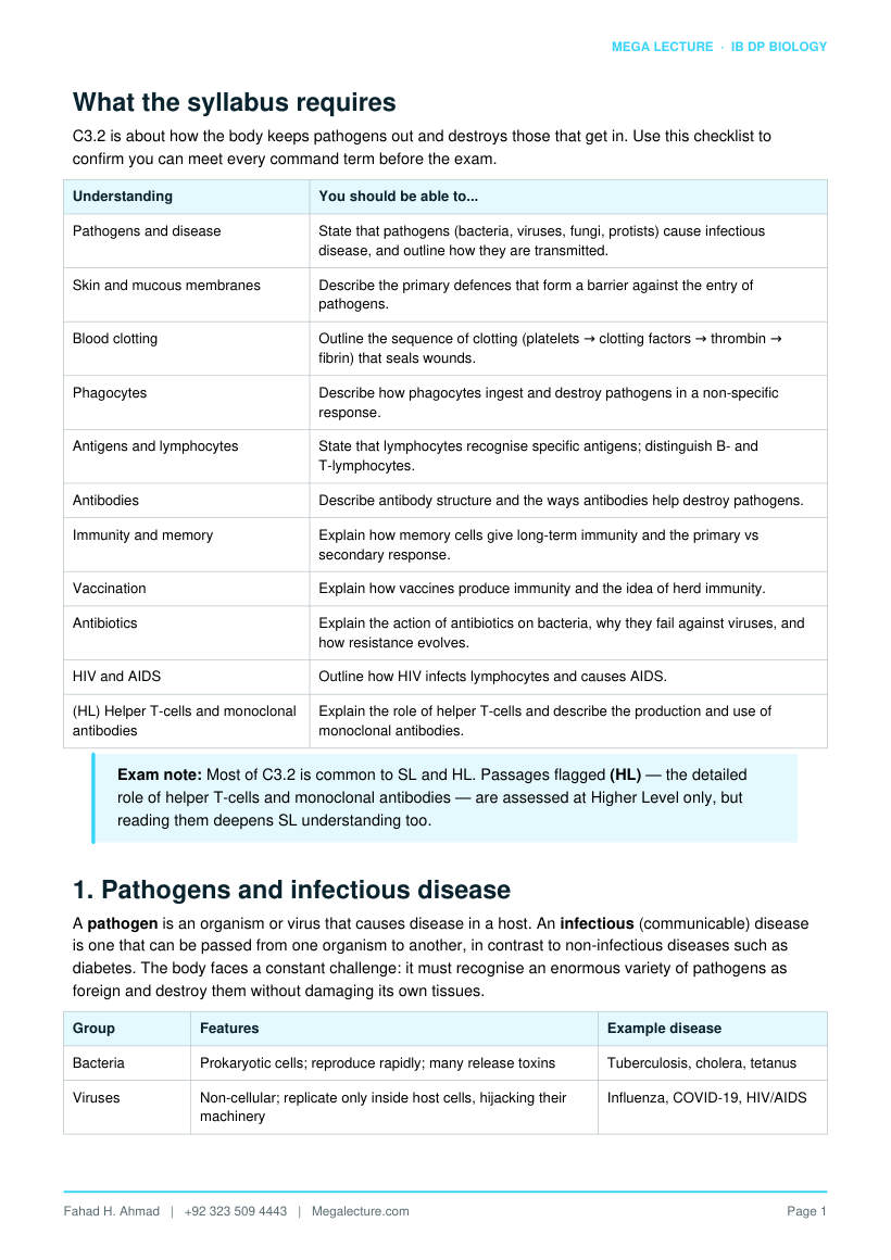
C3.2 is about how the body keeps pathogens out and destroys those that get in. Use this checklist to confirm you can meet every command term before the exam.
Understanding You should be able to...
Pathogens and disease State that pathogens (bacteria, viruses, fungi, protists) cause infectious disease, and outline how they are transmitted.
Skin and mucous membranes Describe the primary defences that form a barrier against the entry of pathogens.
Blood clotting Outline the sequence of clotting (platelets → clotting factors → thrombin → fibrin) that seals wounds.
Phagocytes Describe how phagocytes ingest and destroy pathogens in a non-specific response.
Antigens and lymphocytes State that lymphocytes recognise specific antigens; distinguish B- and T-lymphocytes.
Antibodies Describe antibody structure and the ways antibodies help destroy pathogens.
Immunity and memory Explain how memory cells give long-term immunity and the primary vs secondary response.
Vaccination Explain how vaccines produce immunity and the idea of herd immunity.
Antibiotics Explain the action of antibiotics on bacteria, why they fail against viruses, and how resistance evolves.
HIV and AIDS Outline how HIV infects lymphocytes and causes AIDS.
(HL) Helper T-cells and monoclonal antibodies
Explain the role of helper T-cells and describe the production and use of monoclonal antibodies.
Exam note: Most of C3.2 is common to SL and HL. Passages flagged (HL) — the detailed role of helper T-cells and monoclonal antibodies — are assessed at Higher Level only, but reading them deepens SL understanding too.
1. Pathogens and infectious disease
A pathogen is an organism or virus that causes disease in a host. An infectious (communicable) disease is one that can be passed from one organism to another, in contrast to non-infectious diseases such as diabetes. The body faces a constant challenge: it must recognise an enormous variety of pathogens as foreign and destroy them without damaging its own tissues.
Group Features Example disease
Bacteria Prokaryotic cells; reproduce rapidly; many release toxins Tuberculosis, cholera, tetanus
Viruses Non-cellular; replicate only inside host cells, hijacking their machinery
Influenza, COVID-19, HIV/AIDS
Group Features Example disease
Fungi Eukaryotic; often infect skin or moist surfaces Athlete's foot, ringworm, thrush
Protists Single-celled eukaryotes; often spread by vectors Malaria, sleeping sickness
How pathogens are transmitted
- Direct contact — touching an infected person or contaminated surface (e.g. many fungal and skin infections).
- Droplets in air — coughing and sneezing spread respiratory pathogens (e.g. influenza, tuberculosis).
- Contaminated food and water — the faecal-oral route spreads gut pathogens (e.g. cholera).
- Body fluids — blood or sexual contact (e.g. HIV, hepatitis B).
- Vectors — an animal carrier, such as the mosquito that transmits the malaria protist.
The body's defences are organised in three lines: barriers that keep pathogens out (first line); non-specific innate responses that attack anything foreign (second line); and the specific adaptive immune response that targets a particular pathogen and remembers it (third line).
Command-term tip: “Outline” transmission means giving the route briefly; “describe” a defence means a fuller step-by-step account. Match the detail of your answer to the command term and the marks available.
2. First line of defence: barriers
The first line is non-specific: it resists all pathogens equally and does not target any particular species. Most of it is a physical and chemical barrier at the body surface, preventing entry in the first place.
Barrier How it defends
Skin A tough, dry, keratinised outer layer physically blocks pathogens; its slightly acidic surface discourages microbial growth.
Mucous membranes Line the airways, gut and reproductive tract; sticky mucus traps pathogens, and cilia sweep trapped material away from the lungs.
Stomach acid Hydrochloric acid gives a very low pH that kills most swallowed pathogens.
Lysozyme An enzyme in tears, saliva and mucus that digests bacterial cell walls, causing the bacteria to burst.
Commensal (natural) flora
Harmless bacteria living on skin and in the gut compete with pathogens for space and nutrients, limiting their growth.
Blood clotting seals wounds
A cut breaches the skin, so clotting quickly plugs the gap to stop blood loss and block pathogen entry. The outline sequence is:
- Platelets stick to the exposed, damaged tissue at the wound and release clotting factors (and signalling chemicals).
- These factors trigger a clotting cascade — a chain of reactions in which one activated factor activates the next.
- The cascade converts the inactive enzyme prothrombin into active thrombin.
- Thrombin catalyses the conversion of soluble fibrinogen into insoluble threads of fibrin.
- The fibrin mesh traps platelets and red blood cells, forming a clot that dries into a scab, sealing the wound while new skin grows beneath.
Exam note: You are only expected to outline clotting. Learn the order platelets → clotting factors → thrombin → fibrin → clot, and that fibrin forms the mesh — you do not need every named factor.
3. Second line: innate, non-specific defence
If a pathogen breaches the barriers, the innate response attacks it at once. It is non-specific (the same response to any pathogen) and has no memory. Its main agents are phagocytes, backed by inflammation and fever.
Phagocytes and phagocytosis
Phagocytes are white blood cells that engulf and digest pathogens. The two main types are neutrophils (numerous, fast-acting, short-lived) and macrophages (larger, longer-lived, and also alert the adaptive response). Phagocytosis proceeds in clear steps:
- The phagocyte is attracted to the pathogen by chemicals released at the site of infection (chemotaxis).
- It recognises and binds to the pathogen surface, often more easily when the pathogen is already coated with antibodies.
- The cell membrane wraps around and engulfs the pathogen, enclosing it in a vesicle called a phagosome.
- Lysosomes fuse with the phagosome and release digestive enzymes that break down the pathogen.
- Harmless products are absorbed or expelled; a macrophage may display fragments of the pathogen to activate lymphocytes.
Figure 3. Left: the first-line physical/chemical barrier blocks most pathogens at the body surface. Right: the 4-step phagocytosis sequence used by the non-specific second line once a pathogen breaches the barrier.
Inflammation
Damaged cells and mast cells release histamine, which makes local blood vessels widen (vasodilation) and become more permeable ('leaky'). This increases blood flow to the area — causing the redness, heat, swelling and pain of inflammation — and delivers more phagocytes and clotting factors to fight infection and repair tissue.
Fever
Chemical signals can reset the body's thermostat in the brain to raise core temperature. A moderate fever slows the reproduction of many pathogens and speeds up the immune response, helping the body clear the infection.
Common misconception: Phagocytes do not “recognise a specific pathogen” the way lymphocytes do. They respond to anything identified as foreign — that is what makes the second line non-specific.
4. Third line: adaptive, specific immunity
The adaptive (specific) response targets a particular pathogen and builds memory of it. It is slower to start than the innate response but far more precise, and it is the basis of long-term immunity and vaccination.
Antigens and lymphocytes
An antigen is a molecule (usually a protein or polysaccharide) on the surface of a pathogen that the immune system recognises as foreign. Each pathogen carries its own antigens, allowing it to be identified specifically.
Lymphocytes are white blood cells made in the bone marrow. Each lymphocyte carries receptors for just one specific antigen, so between them the body's lymphocytes can recognise an enormous range of pathogens. Two main types matter here:
- B-lymphocytes (B-cells) — mature in the bone marrow; make antibodies (humoral response).
- T-lymphocytes (T-cells) — mature in the thymus; include helper T-cells that coordinate the response.
The humoral response: clonal selection
The humoral response involves antibodies produced by B-cells and works by clonal selection and clonal expansion:
- A pathogen enters and its antigen is encountered. Only the B-cell whose receptor matches that antigen is selected (clonal selection).
- A helper T-cell that recognises the same antigen binds to the B-cell and releases signals that activate it.
- The activated B-cell divides rapidly by mitosis to form a large clone of identical cells (clonal expansion).
- Most of the clone become plasma cells that secrete large quantities of antibodies specific to that antigen.
- Some of the clone become long-lived memory cells that remain in the body, ready to respond rapidly if the same pathogen returns.
Key idea: “Selection” means the antigen picks out the one matching B-cell from millions already present; “expansion” means that cell then multiplies. The specificity comes first, the numbers come after.
5. Antibodies: structure and action
Antibodies (immunoglobulins) are proteins secreted by plasma cells. Each has a distinctive Y-shape made of four polypeptide chains.
Region Feature and role
Variable region (tips of the Y) Amino-acid sequence differs between antibodies; forms the two antigen-binding sites, each specific to one antigen (complementary shape).
Constant region (stem of the Y) Same within an antibody class; binds to phagocytes and other immune components, and determines how the antibody is used.
Antigen-binding sites Two per antibody, so one antibody can bind two antigens and link pathogens together.
Figure 2. Antibody structure: 2 antigen-binding sites (variable region) per antibody let it link antigens together, while the constant region recruits other immune components. Plasma cells (differentiated B-cells) secrete large numbers of one identical antibody.
How antibodies help destroy pathogens
Antibodies do not usually kill pathogens directly; they mark and disable them so other defences can finish the job:
- Agglutination — because each antibody has two binding sites, antibodies clump many pathogens together into clusters that cannot spread and are easily engulfed.
- Neutralisation — antibodies bind to toxins or to the surface proteins a pathogen uses to enter cells, blocking their action.
- Opsonisation — antibodies coat a pathogen, acting as a flag that makes it far easier for phagocytes to recognise and engulf.
- Marking for destruction — the constant region recruits other immune components that lyse (burst) the pathogen or trigger its destruction.
Common misconception: Antibodies are not cells and do not “eat” pathogens. They are soluble proteins; the actual destruction is done by phagocytes and other mechanisms the antibody recruits.
6. Immunological memory: primary vs secondary response
The first time the body meets a particular antigen it mounts a primary response. Clonal selection and expansion take time, so there is a delay of several days before enough antibodies are made, and their concentration stays relatively low. Meanwhile the person may show symptoms of the disease. Crucially, memory cells are produced and persist for years.
If the same antigen is met again, the secondary response is triggered. Memory cells are already present and recognise the antigen immediately, so they divide and become plasma cells almost at once. Compared with the primary response, the secondary response is:
- Faster — antibodies appear within a day or two instead of a week.
- Larger — a much higher peak concentration of antibody is reached.
- Longer-lasting — antibody levels stay high for longer.
Because antibodies rise so quickly and to such high levels, the pathogen is destroyed before it can multiply enough to cause symptoms — the person is immune. This is the basis of long-term immunity and of vaccination.
Describing the standard graph
A typical exam graph plots antibody concentration (y-axis) against time (x-axis), with arrows marking a first and a second exposure to the same antigen. After the first exposure the curve rises slowly after a lag, reaches a low peak, then falls. After the second exposure the curve rises almost immediately, much more steeply, to a peak several times higher, and declines more slowly. If a different antigen is introduced, only a fresh primary-type curve is produced — proof that memory is antigen-specific.
Figure 1. Primary and secondary antibody responses after a first exposure (day 0) and a second exposure (day 30) to the same antigen. The computed secondary peak is 9.1× the primary peak (131 vs 14 AU), reached 2.4 days sooner after exposure, and stays above half-maximum for 27 d versus 16 d for the primary response.
Exam technique: When asked to compare responses from a graph, quote the three comparisons — faster onset, higher peak, longer duration — and always credit memory cells as the reason. State that memory cells persist and respond rapidly on re-exposure.
7. Vaccination
A vaccine contains antigens from a pathogen in a form that does not cause serious disease — for example a dead or weakened pathogen, a fragment of it, or its surface protein made by other means. Vaccination deliberately gives the immune system a safe first exposure.
How a vaccine gives immunity
- The vaccine introduces the harmless antigen into the body.
- The antigen triggers a primary response: matching B-cells are selected, expand, and form plasma cells and — most importantly — memory cells.
- No serious illness results, because the antigen cannot cause the disease.
- If the real pathogen infects the person later, the memory cells trigger a rapid, large secondary response that destroys it before symptoms develop. The person is immune.
Herd immunity and eradication
When a high proportion of a population is immune, a pathogen can no longer find enough susceptible hosts to spread. This herd immunity protects even the unvaccinated — including babies and people who cannot be vaccinated. If transmission is interrupted worldwide, a disease can be eradicated: smallpox was declared eradicated in 1980 through a global vaccination programme.
Why boosters are needed, and why some pathogens are hard to vaccinate against
- Boosters re-expose the immune system to the antigen, producing another secondary response that raises antibody levels and memory-cell numbers again, extending immunity.
- Antigenic variation — some pathogens change their surface antigens frequently (e.g. the influenza virus), so existing memory cells no longer recognise them and the vaccine must be updated.
- Pathogens that attack the immune system itself (such as HIV) or that have many strains are especially difficult to vaccinate against.
Ethics link: Vaccination raises issues beyond biology — individual choice versus the community benefit of herd immunity, and the fair global distribution of vaccines. Exams may ask you to discuss such implications, not just the mechanism.
8. Antibiotics and antibiotic resistance
Antibiotics are chemicals that kill bacteria (bactericidal) or stop them reproducing (bacteriostatic). They work by targeting features that bacterial cells have but human cells and viruses do not — for example the bacterial cell wall, or bacterial ribosomes and enzymes, which differ from ours. This is why a well-chosen antibiotic harms the pathogen but not the patient.
Why antibiotics do not work against viruses
Viruses are not cells: they have no cell wall, no ribosomes and no metabolism of their own, and they replicate inside the host's cells using the host's machinery. There is nothing bacteria-specific for an antibiotic to attack, so antibiotics have no effect on viral diseases such as colds, influenza or COVID-19.
How antibiotic resistance evolves
Resistance is a clear example of evolution by natural selection:
- Variation: within a bacterial population, random mutations mean a few cells happen to carry a gene giving resistance to an antibiotic.
- Selection pressure: when the antibiotic is used, it kills the non-resistant bacteria but the resistant ones survive.
- Reproduction: the survivors reproduce rapidly, passing the resistance gene to their offspring (and bacteria can also share resistance genes directly).
- Over generations the frequency of resistance rises until most of the population is resistant and the antibiotic no longer works.
Strains such as MRSA (methicillin-resistant Staphylococcus aureus) have become resistant to several antibiotics, making infections hard to treat. Overuse and misuse — including using antibiotics for viral illnesses and not completing a course — speed this process up.
Responsible use of antibiotics
- Prescribe antibiotics only for bacterial infections, never for viral ones.
- Always complete the full prescribed course, even after symptoms improve.
- Reduce non-medical use, such as routine antibiotics in farm animals.
- Develop new antibiotics and use good hygiene to limit the spread of resistant strains.
Exam technique: When explaining resistance, use the language of natural selection: variation → selection pressure → differential survival → inheritance → change in allele frequency. Avoid saying bacteria “try” or “choose” to become resistant.
9. HIV and AIDS
HIV (human immunodeficiency virus) is a virus that infects and destroys helper T-cells — the very cells that coordinate the adaptive immune response. Over years, as helper T-cell numbers fall, the immune system is progressively weakened.
Because helper T-cells are needed to activate B-cells and other lymphocytes, their loss cripples both the antibody response and the wider adaptive response. When immunity becomes so weak that the body can no longer fight off infections, the condition is called AIDS (acquired immunodeficiency syndrome).
Transmission and consequences
- Transmission is through body fluids: unprotected sexual contact, sharing needles, infected blood, and from mother to baby during birth or breastfeeding.
- HIV is not spread by casual contact such as touching, sharing food or insect bites.
- Consequence: with a failing immune system the person suffers opportunistic infections — infections by pathogens that a healthy immune system would normally control — and certain cancers, which are the usual cause of death.
HIV is a good example of why some pathogens are so hard to defend against: by attacking the immune system itself, and by changing its surface antigens rapidly, it evades the very response the body would use against it.
Link: Note the connection between sections 4, 8 and 9: helper T-cells activate B-cells (section 4), so destroying them (HIV) undermines the antibody response, and antibiotics cannot help because HIV is a virus (section 8).
10. (HL) Helper T-cells and monoclonal antibodies
At Higher Level you need more detail on how helper T-cells activate B-cells, and on how identical antibodies can be manufactured for use in medicine and testing.
(HL) The specific role of helper T-cells
Helper T-cells are the coordinators of the adaptive response. Their activation and action follow a defined sequence:
- A macrophage engulfs a pathogen and displays its antigens on the cell surface, acting as an antigen-presenting cell.
- A helper T-cell with a matching receptor binds to the presented antigen and becomes activated, then divides to form a clone (including memory helper T-cells).
- The activated helper T-cell binds to a B-cell that has recognised the same antigen and releases signalling chemicals (cytokines).
- These signals stimulate the B-cell to divide and differentiate into plasma cells and memory cells — so without helper T-cells the antibody response is very weak. (This is exactly why HIV, which destroys helper T-cells, is so damaging.)
(HL) Monoclonal antibodies
Monoclonal antibodies are identical antibodies, all specific to a single antigen, produced from one clone of cells. Production combines the specificity of a B-cell with the immortality of a tumour cell:
- An animal (usually a mouse) is injected with the chosen antigen so that it makes B-cells producing the required antibody.
- These B-cells are removed and fused with fast-dividing tumour cells to form hybridoma cells, which both make the antibody and divide indefinitely.
- A single hybridoma is selected and cultured to form a clone that secretes large amounts of one identical (monoclonal) antibody.
- The antibody is harvested and purified for use.
Uses of monoclonal antibodies
- Pregnancy tests — antibodies on the test strip bind the hormone hCG in urine, producing a coloured line only when hCG is present.
- Diagnosis — detecting specific molecules, pathogens or blood markers, and locating cancer cells that carry particular antigens.
- Therapy — antibodies designed to bind a target on cancer cells or in autoimmune and inflammatory disease, blocking it or delivering a drug precisely to the target cells.
(HL) Exam note: Learn the logic of hybridoma production: B-cell = specificity, tumour cell = unlimited division; fusing them gives an immortal cell line producing one identical antibody. “Mono-clonal” literally means “from one clone.”
11. Skills, pitfalls and quick reference
Worked examples Worked example 1 — sequence phagocytosis
Q. Place the following stages of phagocytosis in the correct order and name the structures involved: digestion by enzymes; engulfing; attraction to the pathogen; binding.
A. (1) Attraction — the phagocyte moves toward chemicals from the site of infection. (2) Binding — it attaches to the pathogen surface (easier if the pathogen is coated with antibodies). (3) Engulfing — the membrane surrounds the pathogen, enclosing it in a phagosome. (4) Digestion — lysosomes fuse with the phagosome and enzymes break the pathogen down.
Mark-scheme tip: name the phagosome and lysosomes to gain the detail marks.
Worked example 2 — reading a primary/secondary response graph
Q. A graph shows antibody level after a first injection of antigen X on day 0 and a second injection of X on day 30. Describe and explain the difference between the two responses.
A. After the first injection (primary response) antibody rises slowly after a lag of several days and reaches a low peak, because matching B-cells must be selected and undergo clonal expansion before plasma cells can secrete antibody.
After the second injection (secondary response) antibody rises much faster, to a higher peak, and stays high for longer, because memory cells made during the primary response recognise antigen X immediately and rapidly form plasma cells. The person shows no symptoms the second time.
Worked example 3 — explain how a vaccine gives immunity
Q. Explain how vaccination against a bacterial disease makes a person immune. [4]
A. The vaccine introduces harmless antigen from the pathogen; this triggers a primary response in which matching B-cells are selected and expand, forming plasma cells and memory cells; no disease results because the antigen is harmless; if the live pathogen infects later, memory cells give a rapid, large secondary response that destroys it before symptoms appear.
Worked example 4 — explain the evolution of antibiotic resistance
Q. Explain how a population of bacteria becomes resistant to an antibiotic. [4]
A. Random mutation produces variation, so a few bacteria carry a resistance allele; using the antibiotic is a selection pressure that kills non-resistant bacteria while the resistant ones survive; the survivors reproduce and pass on the resistance allele; over generations the frequency of the resistance allele increases until the antibiotic is no longer effective (e.g. MRSA).
Common pitfalls
- Writing that antibiotics can cure a cold or flu — they act only on bacteria, not viruses.
- Saying immunity comes from antibodies staying in the blood forever — long immunity comes from memory cells.
- Calling antibodies “cells” — antibodies are proteins made by plasma cells.
- Confusing the second line (phagocytes, non-specific) with the third line (lymphocytes, specific).
- Saying bacteria “want” or “try” to become resistant — resistance arises by chance mutation, then selection.
- Mixing up B-cells (make antibodies) with helper T-cells (coordinate the response).
Quick-reference table
Term One-line meaning
Pathogen Organism or virus that causes disease
Term One-line meaning
First line Non-specific barriers: skin, mucous membranes, stomach acid, lysozyme, flora
Second line Non-specific innate response: phagocytosis, inflammation, fever
Third line Specific adaptive response: lymphocytes, antibodies, memory
Antigen Foreign molecule that is recognised and triggers a specific response
Antibody Y-shaped protein from plasma cells; binds one specific antigen
B-cell / plasma cell Lymphocyte that becomes a plasma cell and secretes antibodies
Helper T-cell Lymphocyte that activates B-cells and coordinates the response
Memory cell Long-lived cell giving a rapid secondary response and immunity
Vaccine Harmless antigen that induces memory without causing disease
Antibiotic Chemical that kills or inhibits bacteria; useless against viruses
(HL) Monoclonal antibody Identical antibodies from one clone (hybridoma); used in tests and therapy
12. Test yourself
Attempt these without notes; full worked answers follow.
- State two ways the skin acts as a barrier to pathogens. [2]
- Outline the sequence of events in blood clotting, ending with the formation of a clot. [4]
- Explain why the second line of defence is described as non-specific, and the third line as specific. [3]
- Describe the structure of an antibody and explain how agglutination helps destroy pathogens. [4]
- Using the idea of memory cells, explain why a person who has had chickenpox rarely catches it again. [3]
- Explain how herd immunity protects people who have not been vaccinated. [3]
- Explain why antibiotics are effective against bacteria but not against viruses. [3]
- (HL) Outline how monoclonal antibodies are produced and give one use. [4]
Answers
- The tough, dry, keratinised outer layer physically blocks entry; its slightly acidic surface (and sebum) discourages the growth of microorganisms. [2]
- Platelets stick to the damaged tissue and release clotting factors → a cascade of reactions is triggered → prothrombin is converted to thrombin → thrombin converts fibrinogen to insoluble fibrin → the fibrin mesh traps blood cells to form a clot that seals the wound. [4]
- The second line (phagocytes, inflammation, fever) attacks any pathogen in the same way, without distinguishing between species, so it is non-specific. The third line uses lymphocytes whose receptors recognise one particular antigen and produces antibodies against that specific pathogen, so it is specific. [3]
- An antibody is a Y-shaped protein with variable regions forming two antigen-binding sites and a constant region. Because it has two binding sites, one antibody can bind two pathogens; many antibodies clump pathogens together (agglutination) so they cannot spread and are more easily engulfed by phagocytes. [4]
- The first infection produces memory cells specific to the chickenpox virus antigen; these persist for years. On re-exposure they trigger a rapid, large secondary response that destroys the virus before it can multiply enough to cause symptoms — so the person is immune. [3]
- When most of a population is immune, an infected person is surrounded by immune individuals, so the pathogen cannot find enough susceptible hosts to spread. Transmission is interrupted, so unvaccinated people are unlikely to meet the pathogen and are protected indirectly. [3]
- Antibiotics target features unique to bacterial cells, such as the cell wall or bacterial ribosomes/enzymes. Viruses are non-cellular, have no such structures, and replicate inside host cells using host machinery, so there is no bacterial target for the antibiotic to act on. [3]
- (HL) A mouse is injected with the antigen and makes B-cells producing the required antibody; these B-cells are fused with tumour cells to form immortal hybridoma cells; one hybridoma is cultured to give a clone secreting identical (monoclonal) antibody, which is then purified. Use: pregnancy testing (detecting hCG), diagnosis, or targeted cancer therapy. [4]
