
IB Diploma · Biology · SL / HL · Theme A: Unity and Diversity
A1.2 Nucleic Acids
Opens inside this page. Prefer your own PDF app? Open the PDF.
Full text of A1.2 Nucleic Acids
IB DP BIOLOGY Theme A: Unity and Diversity A1.2 Nucleic Acids
Revision Notes · Standard and Higher Level Fahad H. Ahmad
+92 323 509 4443 | Megalecture.com
Original notes prepared for the IB Diploma Programme Biology course (first assessment 2025)
What the syllabus requires
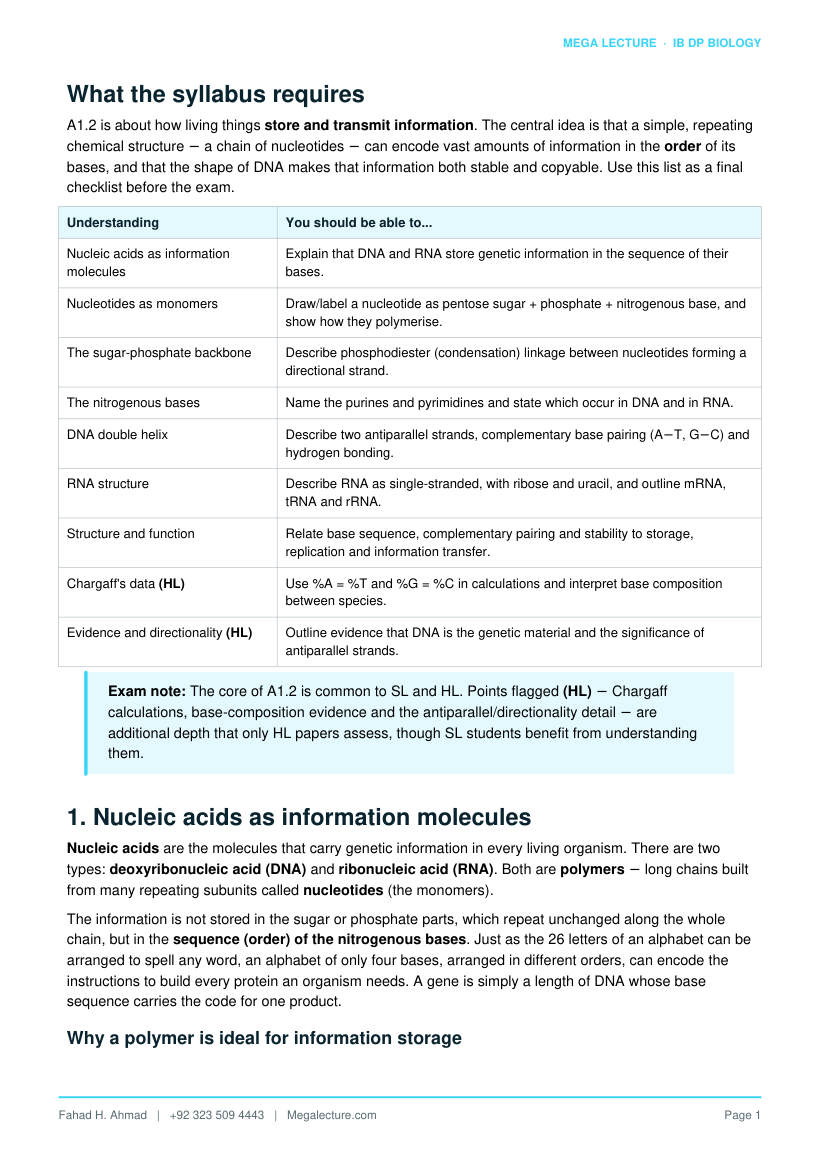
A1.2 is about how living things store and transmit information. The central idea is that a simple, repeating chemical structure − a chain of nucleotides − can encode vast amounts of information in the order of its bases, and that the shape of DNA makes that information both stable and copyable. Use this list as a final checklist before the exam.
Understanding You should be able to...
Nucleic acids as information molecules
Explain that DNA and RNA store genetic information in the sequence of their bases.
Nucleotides as monomers Draw/label a nucleotide as pentose sugar + phosphate + nitrogenous base, and show how they polymerise.
The sugar-phosphate backbone Describe phosphodiester (condensation) linkage between nucleotides forming a directional strand.
The nitrogenous bases Name the purines and pyrimidines and state which occur in DNA and in RNA.
DNA double helix Describe two antiparallel strands, complementary base pairing (A−T, G−C) and hydrogen bonding.
RNA structure Describe RNA as single-stranded, with ribose and uracil, and outline mRNA, tRNA and rRNA.
Structure and function Relate base sequence, complementary pairing and stability to storage, replication and information transfer.
Chargaff's data (HL) Use %A = %T and %G = %C in calculations and interpret base composition between species.
Evidence and directionality (HL) Outline evidence that DNA is the genetic material and the significance of antiparallel strands.
Exam note: The core of A1.2 is common to SL and HL. Points flagged (HL) − Chargaff calculations, base-composition evidence and the antiparallel/directionality detail − are additional depth that only HL papers assess, though SL students benefit from understanding them.
1. Nucleic acids as information molecules
Nucleic acids are the molecules that carry genetic information in every living organism. There are two types: deoxyribonucleic acid (DNA) and ribonucleic acid (RNA). Both are polymers − long chains built from many repeating subunits called nucleotides (the monomers).
The information is not stored in the sugar or phosphate parts, which repeat unchanged along the whole chain, but in the sequence (order) of the nitrogenous bases. Just as the 26 letters of an alphabet can be arranged to spell any word, an alphabet of only four bases, arranged in different orders, can encode the instructions to build every protein an organism needs. A gene is simply a length of DNA whose base sequence carries the code for one product.
Why a polymer is ideal for information storage Where nucleic acids are found, and the scale of the information
In eukaryotic cells the main store of DNA is in the nucleus, wound around proteins and packaged into chromosomes; smaller amounts of DNA are also found in mitochondria and (in plants) chloroplasts. In prokaryotes the DNA is a single circular molecule in the cytoplasm. RNA is made in the nucleus but functions mainly in the cytoplasm, at the ribosomes. The amount of information stored is remarkable: a single human cell contains roughly 3 billion base pairs of DNA, which if unravelled would stretch about two metres, yet it is packed into a nucleus only a few micrometres across. It is the same universal four-base system − A, T (or U), G and C − in every organism on Earth, from bacteria to whales, which is powerful evidence that all life shares a common ancestry.
Key term: A monomer is a single repeating unit; a polymer is a large molecule built by joining many monomers. Nucleotides are the monomers of the nucleic-acid polymers DNA and RNA.
2. The structure of a nucleotide
Every nucleotide is made of three components joined by covalent bonds:
Component Description Role
Pentose sugar A 5-carbon sugar: deoxyribose in DNA, ribose in RNA
Forms the backbone; its carbons are numbered 1′ to 5′
Phosphate group A negatively charged group derived from phosphoric acid
Links sugars together; gives the backbone its acidity
Nitrogenous base One of several ring-shaped, nitrogen-containing molecules
The variable part − its identity carries the information
The sugar sits at the centre. The phosphate group is attached to the 5′ carbon of the sugar, and the nitrogenous base is attached to the 1′ carbon. The carbon numbering (1′ to 5′, read “one-prime” to “five-prime”) matters because it defines the direction of the whole strand, as we shall see.
Figure 1. A single nucleotide: a phosphate group, a pentose (deoxyribose) sugar and a nitrogenous base. The phosphate joins the 5′ carbon; the base joins the 1′ carbon; a free –OH sits on the 3′ carbon.
Joining nucleotides: the sugar-phosphate backbone
Nucleotides are joined by a condensation reaction (a water molecule is removed). The phosphate group on the 5′ carbon of one nucleotide bonds to the 3′ carbon (specifically its –OH group) of the next nucleotide's sugar. This covalent linkage is called a phosphodiester bond.
As nucleotide after nucleotide is added, the alternating sugar−phosphate−sugar−phosphate units form a continuous strand: the sugar-phosphate backbone. The bases project sideways from this backbone. Because the backbone is held together by strong covalent phosphodiester bonds, the sequence of bases is chemically very stable.
Directionality: the 5′ and 3′ ends
A single strand is directional. At one end there is a free phosphate group on a 5′ carbon − this is the 5′ end. At the other end there is a free –OH group on a 3′ carbon − the 3′ end. By convention a base sequence is written and read in the 5′ → 3′ direction, and new nucleotides can only be added to the 3′ end during replication.
Condensation vs hydrolysis: Building the chain (joining nucleotides) is condensation and releases water; breaking the chain is hydrolysis and uses water. The same logic applies to all biological polymers (proteins, polysaccharides, nucleic acids).
Labelling a nucleotide in the exam
A common question shows a nucleotide or a short strand and asks you to label the parts. Look for three things: the phosphate (often drawn as a circle marked P), the pentose sugar (a five-sided ring), and the nitrogenous base (a ring or double ring projecting from the sugar). In a strand, identify the alternating sugar−phosphate backbone and note which end carries the free phosphate (5′) and which the free –OH (3′). Marks are lost by muddling the sugar with the base, or by labelling the whole unit “base” when it is a nucleotide. Practise drawing the three parts as three linked shapes until it is automatic.
3. The nitrogenous bases
There are five nitrogenous bases in total. They fall into two structural groups distinguished by the number of rings in the molecule.
Group Ring structure Bases Found in
Purines Double ring (larger) Adenine (A), Guanine (G) DNA and RNA
Pyrimidines Single ring (smaller) Cytosine (C), Thymine (T), Uracil (U)
C in both; T in DNA only; U in RNA only
So four bases appear in any one nucleic acid:
Adenine, guanine and cytosine are shared by both DNA and RNA. The single difference in the base set is that thymine (DNA) is replaced by uracil (RNA). Thymine and uracil are chemically very similar (thymine is essentially uracil with an extra methyl group), and both pair with adenine.
Memory hooks: PurA-G (purines are Adenine and Guanine, the “pure silver, Ag” pair). CUT the py − Cytosine, Uracil, Thymine are the pyrimidines. A base pair is always one purine + one pyrimidine, keeping the helix a constant width.
4. The structure of DNA
DNA is a double helix: two polynucleotide strands wound around a common axis into a spiral, resembling a twisted ladder. Its structure has several defining features, each with a functional purpose.
Two antiparallel strands
The two strands run in opposite directions − they are antiparallel. One strand runs 5′ → 3′ while its partner runs 3′ → 5′. Where one strand has its 5′ end, the other has its 3′ end. This opposite orientation is essential for the bases to line up and pair correctly.
Backbones outside, bases inside
The two sugar-phosphate backbones lie on the outside of the helix, like the two uprights of a ladder, shielding the information. The nitrogenous bases point inwards towards the centre, forming the rungs of the ladder, where they pair with bases on the opposite strand.
Complementary base pairing
The bases pair according to strict rules: adenine always pairs with thymine (A−T) and guanine always pairs with cytosine (G−C). This is complementary base pairing. Each pair is one purine joined to one pyrimidine, so every rung is the same width and the helix stays uniform.
The two bases in a pair are held together by hydrogen bonds:
Base pair Type of pairing Hydrogen bonds Note
A−T Purine + pyrimidine 2 hydrogen bonds In RNA, A pairs with U (also 2 bonds)
G−C Purine + pyrimidine 3 hydrogen bonds Stronger; more G−C means a more stable helix
Each hydrogen bond is weak on its own, but along a whole molecule there are millions of them, so together they hold the two strands securely. Crucially, they are still weak enough to be “unzipped” by enzymes when the DNA must be copied or read − a perfect compromise between stability and access.
Figure 2. The DNA double helix: two antiparallel sugar-phosphate backbones (one running 5′ → 3′, the other 3′ → 5′) linked by complementary base pairs − A=T (2 H-bonds) and G≡C (3 H-bonds).
The shape of the helix
Because every rung is one purine paired with one pyrimidine, the two backbones stay a constant distance apart, giving the helix a uniform diameter along its whole length. If two purines paired together the rung would be too wide, and two pyrimidines would be too narrow − so the pairing rules are also a geometric requirement, not just a chemical preference. The two strands twist around each other into a right-handed spiral, completing one full turn about every ten base pairs. This coiling makes the molecule compact and helps protect the bases held inside. The regular, repeating shape is the same in the DNA of every organism, which is why a mechanism that copies or reads DNA in one species works on the DNA of another.
Directionality recap: Because the strands are antiparallel, the base at the 5′ end of one strand pairs with the base at the 3′ end of the other. Always keep track of which end is which when writing a complementary strand.
5. The structure of RNA
RNA is also a polymer of nucleotides, but it differs from DNA in three key ways:
The three main types of RNA
RNA molecules do the practical work of expressing the information stored in DNA. Three types are central to protein synthesis (covered in detail in D1):
Type of RNA Full name Role mRNA Messenger RNA Carries a copy of a gene's base sequence from the DNA in the nucleus to the ribosome; its sequence is read to build a protein
Type of RNA Full name Role tRNA Transfer RNA Brings specific amino acids to the ribosome and matches them to the mRNA code through its anticodon rRNA Ribosomal RNA A structural and catalytic component of ribosomes, the organelles where proteins are assembled
The single-stranded, less-stable nature of RNA suits its role as a short-lived working copy of genetic information, whereas the stable double helix of DNA suits long-term storage of the master copy.
Sugar spotting: The name says it all: deoxyribose in DNA is ribose “minus an oxygen”. Ribose (RNA) has the full set of –OH groups; deoxyribose (DNA) lacks the oxygen on the 2′ carbon, which is one reason DNA is the more stable, long-term molecule.
6. DNA versus RNA − summary comparison
This comparison is a very common exam question. Learn it as a table.
Feature DNA RNA
Pentose sugar Deoxyribose Ribose
Bases used A, G, C, T (thymine) A, G, C, U (uracil)
Number of strands Two (double helix) One (single-stranded)
Pairing partner of A Thymine (T) Uracil (U)
Relative stability More stable (long-term) Less stable (short-lived)
Typical length Very long (whole chromosomes) Much shorter (single genes/products)
Main function Stores genetic information as the master copy
Expresses information: makes proteins (mRNA, tRNA, rRNA)
Shared features: Do not forget what DNA and RNA have in common: both are polymers of nucleotides; both use a pentose sugar, a phosphate group and nitrogenous bases; both have a sugar-phosphate backbone; and both share the bases adenine, guanine and cytosine.
7. How structure enables function
The whole point of A1.2 is to link the structure of nucleic acids to what they do. Three structural features explain three functions.
(a) Base sequence stores information
Because any base can follow any other along a strand, the order of bases can vary without limit. This variable sequence is a code: a specific order of bases specifies a specific order of amino acids in a protein, and hence the organism's characteristics. The uniform backbone carries this variable message without altering it.
(b) Complementary pairing enables accurate copying and transfer
Because A only pairs with T and G only with C, each strand carries all the information needed to rebuild the other. During replication the two strands separate and each acts as a template; free nucleotides pair with the exposed bases, producing two identical DNA molecules. The same complementary principle allows information to be transferred to mRNA during transcription. This is why DNA can be copied with such high fidelity from generation to generation.
(c) Stability suits long-term storage
Several features make DNA a robust archive. The covalent sugar-phosphate backbone is strong; the reactive bases are tucked inside the helix, protected by the backbones outside; the double-stranded structure means a damaged base on one strand can be repaired using the intact complementary strand; and DNA lacks the extra 2′ oxygen of RNA, making it chemically more stable. Together these allow DNA to store genetic information reliably for the whole life of a cell and to be passed on unchanged.
The big link: Sequence = information; pairing = copying and transfer; double helix = stability. If you can state each structural feature and the function it serves, you have the core of this topic.
8. Nucleic acids, unity and diversity
Theme A asks you to see both the unity and the diversity of life. Nucleic acids illustrate both at once, which is why this topic sits at the start of the course.
Unity: one shared molecular language
Every known organism stores its genetic information in nucleic acids built from the same handful of nucleotides, joined by the same phosphodiester backbone, and read in the same 5′ → 3′ direction. In almost all organisms the same three-base codons specify the same amino acids − the genetic code is essentially universal. This deep similarity is strong evidence that all life descended from a common ancestor, and it is what makes genetic engineering possible: a human gene inserted into a bacterium can still be read and translated, because both use the same molecular language.
Diversity: endless variation in one sequence
At the same time, nucleic acids are the source of the diversity of life. Because the four bases can be arranged in any order along a chain of millions of units, the number of possible sequences is astronomically large. Differences in base sequence between individuals and species account for the differences in their proteins and therefore their characteristics. Changes to the sequence (mutations) create new variation, which is the raw material for evolution. So the same class of molecule provides both the sameness that unites all living things and the variation that makes each species − and each individual − unique.
Theme link: One molecular system, endless sequences: the structure of nucleic acids is shared by all life (unity), while the sequence of bases differs between all life (diversity). This is the heart of Theme A.
9. Chargaff's rules (HL)
(HL) Before the double helix was known, Erwin Chargaff measured the amounts of each base in DNA from many species and found a consistent pattern, now called Chargaff's rules:
These equalities are a direct consequence of complementary base pairing: every A on one strand is matched by a T on the other, and every G by a C. It follows that the total amount of purines equals the total amount of pyrimidines:
A + G = T + C (purines = pyrimidines, ratio 1 : 1) Using Chargaff's rules in calculations
Since the four base percentages must add up to 100%, knowing just one value lets you find all four. The method is:
- If you know %A, then %T = %A (they are equal). 2. The remaining percentage, 100% − (%A + %T), is shared equally by G and C. 3. So %G = %C = [100% − (%A + %T)] ÷ 2.
Base composition as evidence
While %A = %T and %G = %C hold for every organism, the ratio of (A+T) to (G+C) differs between species. Human DNA, a bacterium and a plant each have a characteristic base composition. This variation was early evidence that DNA carries species-specific information, and that the differences between organisms lie in the sequence and proportion of bases rather than in the backbone. (Note that two unrelated species can share a similar overall %GC yet still have completely different base sequences − composition and sequence are not the same thing.)
Figure 3. Left: A pairs with T by 2 hydrogen bonds, G pairs with C by 3 hydrogen bonds. Right: applying Chargaff to the worked example − given %A = 28%, then %T = 28% and %G = %C = 22%.
HL exam tip: Chargaff's equalities apply to double-stranded DNA. They do not hold for a single strand on its own, nor for single-stranded RNA − a favourite trap. If a question gives you one strand only, %A need not equal %T on that strand.
10. DNA as the genetic material and the meaning of antiparallel (HL)
(HL) That DNA − rather than protein − is the hereditary molecule was established by a series of classic experiments. You are not expected to recall every detail, but you should understand the logic of the evidence.
Transformation evidence
Early work with bacteria showed that a harmless strain could be permanently changed (“transformed”) into a disease-causing strain by something released from dead cells of the harmful strain. When the different chemical components were separated and tested, the transforming factor was shown to be DNA: destroying the DNA abolished the effect, whereas destroying proteins did not. This pointed to DNA as the carrier of inherited information.
Viral (bacteriophage) evidence
A virus that infects bacteria (a bacteriophage) is made of only DNA and a protein coat. By labelling the DNA and the protein separately, experiments showed that it is the phage's DNA that enters the bacterium and directs the production of new viruses, while the protein coat stays outside. Since only the DNA went in and carried the instructions, DNA must be the genetic material. Together these lines of evidence convinced biologists that genes are made of DNA.
The significance of antiparallel strands
The antiparallel arrangement is not a trivial detail − it is required for the molecule to work:
Directionality in one line: Antiparallel means “parallel but pointing opposite ways”: 5′ → 3′ on one strand sits alongside 3′ ← 5′ on the other. It is what makes complementary pairing and accurate copying possible.
11. Worked examples
Worked example 1 − Chargaff calculation
A sample of double-stranded DNA is found to contain 28% adenine. Calculate the percentage of each of the other three bases. (HL)
Step 1: %A = %T, so %T = 28%.
Step 2: %A + %T = 28 + 28 = 56%. The rest, 100 − 56 = 44%, is shared by G and C.
Step 3: %G = %C = 44 ÷ 2 = 22% each.
Answer: A = 28%, T = 28%, G = 22%, C = 22%. Check: 28 + 28 + 22 + 22 = 100%. ✓
Worked example 2 − writing a complementary strand
One strand of DNA reads 5′−A T G C G T A C−3′. Write the sequence of the complementary strand, showing its polarity.
Replace each base with its partner (A−T, G−C) and remember the new strand is antiparallel:
Template: 5′−A T G C G T A C−3′
Partner: 3′−T A C G C A T G−5′
So the complementary strand, written conventionally 5′ → 3′, is 5′−G T A C G C A T−3′.
Worked example 3 − counting hydrogen bonds
A short double-stranded DNA segment is 20 base pairs long and contains 12 A−T pairs. How many hydrogen bonds hold the two strands together?
A−T pairs: 12, each with 2 hydrogen bonds → 12 × 2 = 24 bonds.
G−C pairs: 20 − 12 = 8, each with 3 hydrogen bonds → 8 × 3 = 24 bonds.
Total = 24 + 24 = 48 hydrogen bonds. Note that a G−C-rich region needs more energy to separate because it has more hydrogen bonds.
Worked example 4 − identifying 5′ and 3′ ends
How can you tell the 5′ end of a strand from the 3′ end, and why does it matter?
The 5′ end has a free phosphate group attached to the 5′ carbon of the terminal sugar.
The 3′ end has a free –OH (hydroxyl) group on the 3′ carbon of the terminal sugar.
It matters because sequences are read 5′ → 3′, the two strands are antiparallel, and enzymes add new nucleotides only to the 3′ end.
Worked example 5 − purine : pyrimidine ratio
In a sample of double-stranded DNA, adenine makes up 31% of the bases. Show that the ratio of purines to pyrimidines is 1 : 1. (HL)
From Chargaff: %T = %A = 31%. Remaining 100 − 62 = 38% is G + C, so %G = %C = 19%.
Purines = %A + %G = 31 + 19 = 50%. Pyrimidines = %T + %C = 31 + 19 = 50%.
Ratio = 50 : 50 = 1 : 1. This is always true in double-stranded DNA, because every purine on one strand is paired with a pyrimidine on the other.
Worked example 6 − interpreting base-composition data
A nucleic acid sample is analysed and found to contain A = 30%, G = 20%, C = 20%, U = 30%, and no thymine. Identify the type of nucleic acid and state whether it is single- or double-stranded, giving reasons.
It contains uracil and no thymine, so it must be RNA (thymine occurs only in DNA).
Here %A = %U and %G = %C only by coincidence of the numbers; RNA is normally single-stranded, so Chargaff-style equalities are not required and cannot be assumed. Because it is RNA, we conclude it is single-stranded.
Answer: single-stranded RNA. Note the trap: equal %A/%U here does not prove double-stranded − that reasoning applies only to double-stranded DNA.
12. Common exam pitfalls 13. Quick reference
Item Key fact
Monomer Nucleotide = pentose sugar + phosphate group + nitrogenous base
Backbone bond Phosphodiester bond (formed by condensation, 5′ phosphate to 3′ –OH)
Purines Adenine (A), Guanine (G) − double ring
Pyrimidines Cytosine (C), Thymine (T), Uracil (U) − single ring
DNA bases A, G, C, T
RNA bases A, G, C, U (uracil replaces thymine)
Base pairing A−T (2 H-bonds), G−C (3 H-bonds); A−U in RNA
DNA shape Antiparallel double helix; backbones outside, bases inside
RNA shape Single-stranded; ribose sugar
Chargaff (HL) %A = %T and %G = %C in double-stranded DNA; purines = pyrimidines
Directionality Read 5′ → 3′; nucleotides added to the 3′ end
14. Test yourself
Attempt these without notes; full answers follow.
- List the three components of a nucleotide and name the bond that joins one nucleotide to the next. (3)
- State the base-pairing rules in DNA and give the number of hydrogen bonds in each pair. (3)
- Explain what is meant by “antiparallel” and why it is important. (3)
- Give three differences between DNA and RNA. (3)
- Explain how the structure of DNA makes it suitable for the long-term storage of genetic information. (3)
- (HL) A sample of double-stranded DNA contains 20% cytosine. Calculate the percentage of adenine, thymine and guanine. (3)
- (HL) A double-stranded DNA molecule has 15 A−T base pairs and 10 G−C base pairs. How many hydrogen bonds hold it together? (2)
- One strand of DNA reads 5′−T A C G G A T−3′. Write the complementary strand in the correct orientation, and write the mRNA sequence that would be transcribed from this strand. (3)
Answers
- A pentose sugar, a phosphate group and a nitrogenous base (1+1 for all three); nucleotides are joined by a phosphodiester bond, formed by condensation (1).
- Adenine pairs with thymine (A−T) with 2 hydrogen bonds (1+1); guanine pairs with cytosine (G−C) with 3 hydrogen bonds (1).
- The two strands run in opposite directions − one 5′ → 3′ and the other 3′ → 5′ (1). This is required so that the complementary bases can align and form hydrogen bonds correctly (1), and it determines how the strands are copied during replication (1).
- Any three: DNA has deoxyribose, RNA has ribose; DNA uses thymine, RNA uses uracil; DNA is double-stranded, RNA is single-stranded; DNA is longer/more stable, RNA is shorter/less stable (1 each, max 3).
- The sugar-phosphate backbone is held by strong covalent bonds (1); the reactive bases are protected on the inside of the helix, and the double-stranded structure allows damage to one strand to be repaired using the other (1); the base sequence stores information stably and can be copied accurately by complementary base pairing (1).
- %C = 20%, so %G = 20% (%G = %C) (1). A + T = 100 − (20 + 20) = 60%, shared equally, so %A = %T = 30% each (1+1). A = 30%, T = 30%, G = 20%.
- A−T: 15 × 2 = 30 bonds; G−C: 10 × 3 = 30 bonds (1); total = 60 hydrogen bonds (1).
- Complementary DNA strand (antiparallel): 3′−A T G C C T A−5′, i.e. written 5′ → 3′ it is 5′−A T C C G T A−3′ (1+1). The mRNA transcribed from the given strand uses U in place of T and A−U pairing: 3′−A U G C C U A−5′ (1).
