
GCSE · Biology · Cell biology: revision notes and key terms
Cell biology: revision notes and key terms
Opens inside this page. Prefer your own PDF app? Open the PDF.
Check yourself
5 quick questions on GCSE Biology
Tap an answer to see if you are right, and why. Free, no sign-up.
Start the free testWas this note useful?
Full text of Cell biology: revision notes and key terms
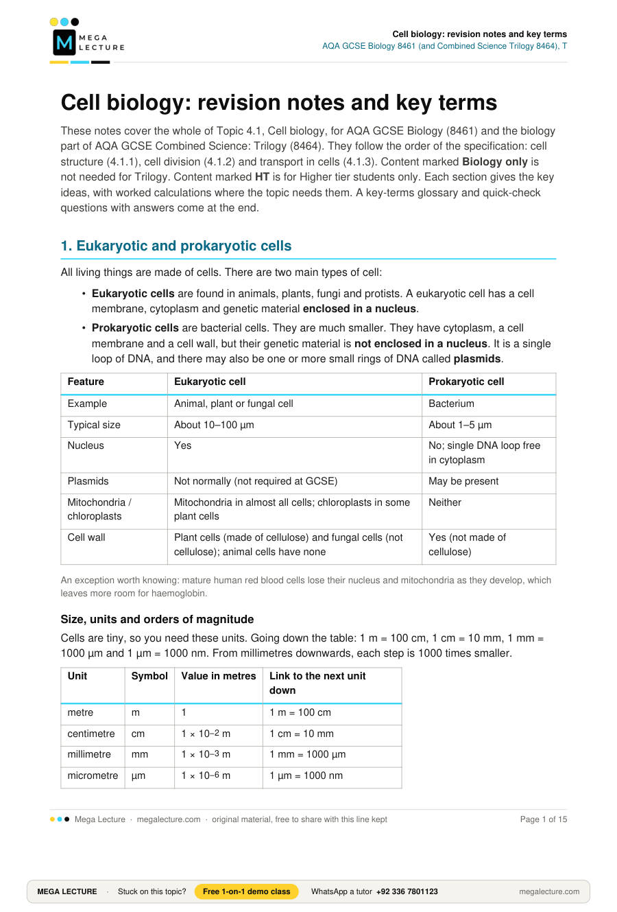
These notes cover the whole of Topic 4.1, Cell biology, for AQA GCSE Biology (8461) and the biology part of AQA GCSE Combined Science: Trilogy (8464). They follow the order of the specification: cell structure (4.1.1), cell division (4.1.2) and transport in cells (4.1.3). Content marked Biology only is not needed for Trilogy. Content marked HT is for Higher tier students only. Each section gives the key ideas, with worked calculations where the topic needs them. A key-terms glossary and quick-check questions with answers come at the end.
1. Eukaryotic and prokaryotic cells
All living things are made of cells. There are two main types of cell:
- Eukaryotic cells are found in animals, plants, fungi and protists. A eukaryotic cell has a cell membrane, cytoplasm and genetic material enclosed in a nucleus.
- Prokaryotic cells are bacterial cells. They are much smaller. They have cytoplasm, a cell membrane and a cell wall, but their genetic material is not enclosed in a nucleus. It is a single loop of DNA, and there may also be one or more small rings of DNA called plasmids.
| Feature | Eukaryotic cell | Prokaryotic cell |
|---|---|---|
| Example | Animal, plant or fungal cell | Bacterium |
| Typical size | About 10–100 µm | About 1–5 µm |
| Nucleus | Yes | No; single DNA loop free in cytoplasm |
| Plasmids | Not normally (not required at GCSE) | May be present |
| Mitochondria / chloroplasts | Mitochondria in almost all cells; chloroplasts in some plant cells | Neither |
| Cell wall | Plant cells (made of cellulose) and fungal cells (not cellulose); animal cells have none | Yes (not made of cellulose) |
An exception worth knowing: mature human red blood cells lose their nucleus and mitochondria as they develop, which leaves more room for haemoglobin.
Size, units and orders of magnitude
Cells are tiny, so you need these units. Going down the table: 1 m = 100 cm, 1 cm = 10 mm, 1 mm = 1000 µm and 1 µm = 1000 nm. From millimetres downwards, each step is 1000 times smaller.
| Unit | Symbol | Value in metres | Link to the next unit down |
|---|---|---|---|
| metre | m | 1 | 1 m = 100 cm |
| centimetre | cm | 1 × 10⁻² m | 1 cm = 10 mm |
| millimetre | mm | 1 × 10⁻³ m | 1 mm = 1000 µm |
| micrometre | µm | 1 × 10⁻⁶ m | 1 µm = 1000 nm |
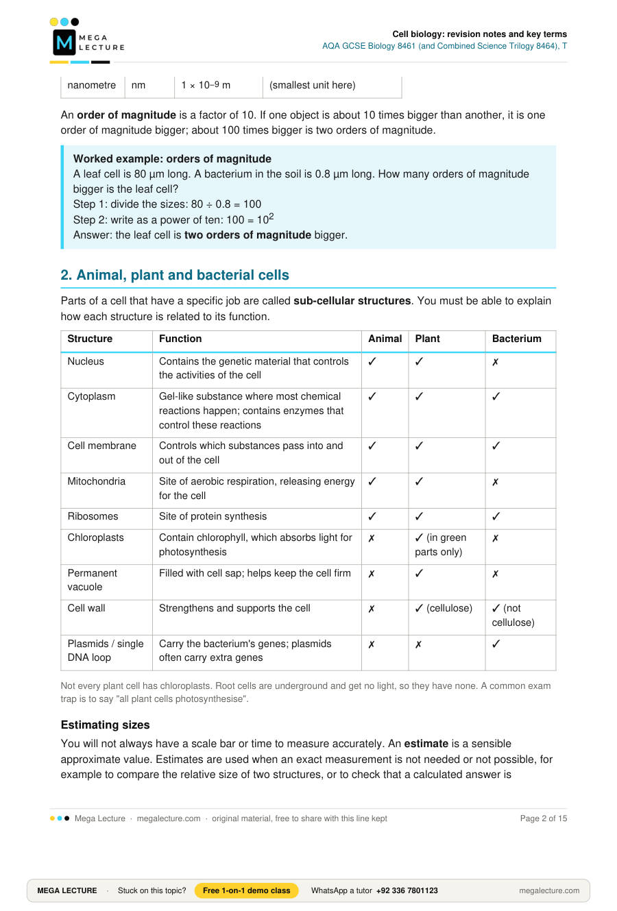
| nanometre | nm | 1 × 10⁻⁹ m | (smallest unit here) |
An order of magnitude is a factor of 10. If one object is about 10 times bigger than another, it is one order of magnitude bigger; about 100 times bigger is two orders of magnitude.
Worked example: orders of magnitudeA leaf cell is 80 µm long. A bacterium in the soil is 0.8 µm long. How many orders of magnitude bigger is the leaf cell?
Step 1: divide the sizes: 80 ÷ 0.8 = 100
Step 2: write as a power of ten: 100 = 102
Answer: the leaf cell is two orders of magnitude bigger.
2. Animal, plant and bacterial cells
Parts of a cell that have a specific job are called sub-cellular structures. You must be able to explain how each structure is related to its function.
| Structure | Function | Animal | Plant | Bacterium |
|---|---|---|---|---|
| Nucleus | Contains the genetic material that controls the activities of the cell | ✓ | ✓ | ✗ |
| Cytoplasm | Gel-like substance where most chemical reactions happen; contains enzymes that control these reactions | ✓ | ✓ | ✓ |
| Cell membrane | Controls which substances pass into and out of the cell | ✓ | ✓ | ✓ |
| Mitochondria | Site of aerobic respiration, releasing energy for the cell | ✓ | ✓ | ✗ |
| Ribosomes | Site of protein synthesis | ✓ | ✓ | ✓ |
| Chloroplasts | Contain chlorophyll, which absorbs light for photosynthesis | ✗ | ✓ (in green parts only) | ✗ |
| Permanent vacuole | Filled with cell sap; helps keep the cell firm | ✗ | ✓ | ✗ |
| Cell wall | Strengthens and supports the cell | ✗ | ✓ (cellulose) | ✓ (not cellulose) |
| Plasmids / single DNA loop | Carry the bacterium's genes; plasmids often carry extra genes | ✗ | ✗ | ✓ |
Not every plant cell has chloroplasts. Root cells are underground and get no light, so they have none. A common exam trap is to say "all plant cells photosynthesise".
Estimating sizes
You will not always have a scale bar or time to measure accurately. An estimate is a sensible approximate value. Estimates are used when an exact measurement is not needed or not possible, for example to compare the relative size of two structures, or to check that a calculated answer is sensible.
Worked example: estimating the size of a nucleusOn a micrograph, the nucleus of a cheek cell looks about one-tenth as wide as the whole cell. The cell is 50 µm wide.
Step 1: estimate the fraction: nucleus ≈ 1/10 of the cell's width
Step 2: 50 µm ÷ 10 = 5 µm
Answer: the nucleus is about 5 µm across. By the same method you could estimate that the nucleus takes up roughly 1/100 of the area of the cell's outline (1/10 × 1/10), because area depends on width × height.
3. Cell specialisation and differentiation
Cells in multicellular organisms become specialised: their structure is adapted to one particular function.
| Cell | Function | Adaptations |
|---|---|---|
| Sperm cell | Reach and fertilise an egg | Tail to swim; many mitochondria to release energy for swimming; enzymes in the head to digest a way into the egg; nucleus with half the normal number of chromosomes |
| Nerve cell | Carry electrical impulses around the body | Long axon to carry impulses over a long distance; many branched endings to connect with other nerve cells |
| Muscle cell | Contract to bring about movement | Contains protein fibres that slide to shorten the cell; many mitochondria to release energy for contraction |
| Root hair cell | Absorb water and mineral ions from soil | Long hair-like projection gives a large surface area; no chloroplasts; many mitochondria to release energy for active transport |
| Xylem cell | Carry water and mineral ions up the plant | Cells are dead and hollow with no end walls, forming continuous tubes; walls strengthened with lignin |
| Phloem cell | Carry dissolved sugars around the plant | Sieve tube cells have perforated end walls (sieve plates) so sap can flow; few sub-cellular structures; companion cells supply energy |
Every cell in a plant or animal comes from one fertilised egg. At first the new cells all look alike. Differentiation is the change that turns an unspecialised cell into a specialised one, as it picks up the sub-cellular structures its job needs.
Example: a cell made in the meristem at a shoot tip can become part of a xylem vessel. It grows longer, lignin builds up in its walls, and then it dies and its end walls break down, leaving a hollow tube for water.
Animals and plants differ here. In animals, most cells lose the ability to differentiate early in development, so in an adult, cell division is mostly about repairing damage and replacing worn-out cells. Many plant cells can still differentiate throughout the plant's life.
4. Microscopy (Required Practical 1)
Light microscopes use light and lenses. They are cheap, portable and can show living cells. Their resolution is enough to see nuclei, cell walls, vacuoles and chloroplasts, but not ribosomes or the detail inside mitochondria. Electron microscopes use a beam of electrons. They have much higher magnification and resolving power, so they show sub-cellular structures such as mitochondria and ribosomes in fine detail.
How microscopy has developed
Simple light microscopes in the 1600s let scientists see cells for the first time; Robert Hooke used the word "cells" after looking at thin slices of cork in 1665. Better lenses in the 1800s made nuclei visible and helped show that all living things are made of cells. The electron microscope was developed in the 1930s. Because its resolution is so much higher, it revealed ribosomes, the folded inner membranes of mitochondria and the internal structure of chloroplasts. This has greatly increased our understanding of sub-cellular structures and how they work.
| Light microscope | Electron microscope | |
|---|---|---|
| Maximum useful magnification | About ×1500–2000 | Up to about ×2 000 000 |
| Resolution | About 200 nm | Down to about 0.1–0.2 nm |
| Living specimens? | Yes | No (specimen in a vacuum) |
| Cost and size | Cheap, small | Very expensive, large |
Magnification: how many times bigger the image is than the real object.
Resolution (resolving power): the ability to distinguish between two points that are close together; higher resolution gives a sharper, more detailed image.
Magnification = size of image ÷ size of real object
Rearranged: size of real object = size of image ÷ magnification. Always convert both sizes into the same unit first. Magnification has no unit, but is often written with a × sign.
Worked example 1: finding real sizeIn a photo taken with a light microscope, a cheek cell is 36 mm wide. The magnification is ×600. Calculate the real width of the cell in µm.
Step 1: real size = image size ÷ magnification = 36 mm ÷ 600 = 0.06 mm
Step 2: convert to µm (× 1000): 0.06 × 1000 = 60 µm
Step 3 (standard form, in metres): 60 µm = 60 × 10−6 m = 6.0 × 10⁻⁵ m Worked example 2: finding magnification
A mitochondrion is 1.5 µm long. On an electron micrograph it measures 45 mm. Calculate the magnification.
Step 1: same units: 45 mm = 45 × 1000 = 45 000 µm
Step 2: magnification = image size ÷ real size = 45 000 ÷ 1.5 = 30 000
Answer: ×30 000 (3 × 10⁴) Worked example 3: using a scale bar
A scale bar on a drawing is labelled 20 µm and measures 8 mm with a ruler.
Step 1: 8 mm = 8000 µm
Step 2: magnification = 8000 ÷ 20 = ×400
You can now use ×400 to find the real size of anything else on the drawing.
Required Practical 1: using a light microscope
- Place a thin specimen (for example, a single layer of onion epidermis) on a slide in a drop of water; add a stain such as iodine solution to make structures visible. Lower a coverslip at an angle to avoid air bubbles.
- Clip the slide onto the stage and switch on the lamp (or adjust the mirror).
- Start with the lowest-power objective lens. Use the coarse focus to bring the stage close to the lens while watching from the side, then focus by moving it away.
- Use the fine focus to sharpen the image, then switch to a higher-power objective and refocus with the fine focus.
- Total magnification = eyepiece lens magnification × objective lens magnification (for example, ×10 × ×40 = ×400).
- Draw with a sharp pencil, using clear continuous lines, no shading, labels with ruled lines, and include the magnification or a scale bar.
5. Culturing microorganisms (Biology only)
Bacteria reproduce by splitting in two, a process called binary fission. With plenty of nutrients and a suitable temperature, some bacteria can divide about once every 20 minutes. In the lab they are grown either in a liquid nutrient broth or on the surface of an agar gel plate, where each original cell grows into a visible colony. To test how well a disinfectant or antibiotic works, the culture must contain only the bacteria being studied; if other microorganisms get in, you cannot tell which organism the chemical affected.
Aseptic technique and why each step matters
- Petri dishes and culture media are sterilised before use (for example, in an autoclave) to kill any microorganisms already present.
- Inoculating loops are sterilised by passing them through a Bunsen flame until red hot, then allowed to cool so they do not kill the bacteria being transferred.
- The lid of the Petri dish is secured with adhesive tape to stop microorganisms from the air getting in. Only a few short strips are used, not a seal all round, so that oxygen can still get in; sealing it completely would create anaerobic conditions that favour harmful anaerobic bacteria. The dish is stored upside down so condensation does not drip onto the agar.
- In school laboratories cultures are incubated at a maximum of 25 °C, which greatly reduces the chance of growing pathogens that thrive at body temperature (37 °C).
Calculating bacterial population growth
Each division doubles the number of bacteria, so:
number of divisions = total time ÷ mean division timebacteria at the end = bacteria at the start × 2number of divisions Worked example 4 (Biology only; giving the answer in standard form is HT)
A broth starts with 500 bacteria. The mean division time is 20 minutes. Calculate the number of bacteria after 3 hours, assuming all survive. Give your answer in standard form.
Step 1: convert time: 3 hours = 3 × 60 = 180 minutes
Step 2: number of divisions = 180 ÷ 20 = 9
Step 3: 29 = 512
Step 4: 500 × 512 = 256 000
Step 5 (HT): standard form: 2.56 × 10⁵ bacteria
Required Practical 2: effect of antiseptics or antibiotics
Paper discs soaked in different antiseptics are placed on an agar plate spread with bacteria. After incubation, a clear zone of inhibition appears around effective discs. The bigger the clear zone, the more effective the substance. A disc soaked in sterile water acts as the control. The area of a circular zone is found using area = πr2.
Worked example 5A clear zone has a diameter of 14 mm. Calculate its area.
Step 1: radius = diameter ÷ 2 = 14 ÷ 2 = 7 mm
Step 2: area = π × 72 = π × 49 = 153.9…
Answer: 154 mm2 (3 significant figures)
6. Chromosomes, mitosis and the cell cycle
The nucleus of a cell contains chromosomes made of DNA. Each chromosome carries a large number of genes. In body cells, chromosomes are normally found in pairs (humans have 23 pairs, 46 in total).
A cell that is going to divide works through a repeating sequence called the cell cycle: first it doubles its genetic material, then it shares it out equally so that two genetically identical cells are made.
| Stage | What happens |
|---|---|
| 1. Growth and DNA replication | Before dividing, the cell gets bigger and makes extra sub-cellular structures, such as ribosomes and mitochondria, so each new cell will have enough. Every chromosome is copied, so the cell now holds two copies of each one. |
| 2. Mitosis | The two copies of each chromosome separate. One complete set moves to each end of the cell, and the nucleus divides. |
| 3. Division of cytoplasm | The cytoplasm and cell membrane split, giving two daughter cells that are genetically identical to each other and to the parent cell. |
Cell division by mitosis is important in the growth and development of multicellular organisms, and in repairing damaged tissue and replacing cells. In a micrograph, cells in mitosis are recognised because their chromosomes are visible as dark, separate threads rather than an evenly stained nucleus.
Worked example 6: time spent in mitosis (extension: not a specification requirement, but good practice for percentage and proportion questions)In a root tip, a student counts 250 cells; 20 of them are in mitosis. One full cell cycle takes 16 hours. Estimate the time spent in mitosis.
Step 1: percentage in mitosis = 20 ÷ 250 × 100 = 8%
Step 2: time = 0.08 × 16 hours = 1.28 hours
Step 3: in minutes: 1.28 × 60 = 76.8 ≈ 77 minutes
This works because the proportion of cells seen in a stage reflects the proportion of the cycle spent in it.
7. Stem cells
Learn this: stem cellA stem cell is an undifferentiated cell of an organism which is capable of giving rise to many more cells of the same type, and from which certain other cells can arise by differentiation.
| Source | What it can become | Notes |
|---|---|---|
| Human embryos | Most types of human cell | Can be cloned and made to differentiate; raise ethical issues because the embryo is destroyed |
| Adult bone marrow | Many types of cell, including blood cells | Fewer ethical issues but less versatile |
| Plant meristems (root and shoot tips) | Any type of plant cell | Keep this ability throughout the life of the plant |
Uses
- Stem cells may help conditions such as diabetes (making new insulin-producing cells) and paralysis (making new nerve cells).
- In therapeutic cloning an embryo is produced with the same genes as the patient (the nucleus from one of the patient's body cells is put into an egg cell that has had its nucleus removed). Stem cells from this embryo are not rejected by the patient's body, so they can be used for medical treatment.
- Meristem stem cells can be used to produce clones of plants quickly and economically: to protect rare species from extinction, and to produce large numbers of identical crop plants with useful features such as disease resistance.
Risks and ethical issues
- Stem cells grown in a lab may be contaminated, so a viral infection could be transferred to the patient.
- Some people object on ethical or religious grounds, because embryos that could develop into a person are destroyed.
- Others argue that embryos used for research are often spare embryos from fertility treatment that would be destroyed anyway, and that the potential to relieve suffering is great. In therapeutic cloning, however, an embryo is created specially, so this argument does not apply to it.
"Evaluate" questions on stem cells need points on both sides and then a justified conclusion.
8. Diffusion
Learn this: diffusionDiffusion is the spreading out of the particles of any substance in solution, or particles of a gas, resulting in a net movement from an area of higher concentration to an area of lower concentration. It is passive: it does not need energy from respiration.
Examples in living things: oxygen and carbon dioxide move by diffusion in gas exchange in the lungs and leaves; the waste product urea diffuses from cells into the blood plasma to be removed by the kidney.
Factors that increase the rate of diffusion:
- a bigger difference in concentration (a steeper concentration gradient)
- a higher temperature (particles have more kinetic energy and move faster)
- a larger surface area of the membrane
9. Surface area to volume ratio and exchange surfaces
A single-celled organism has a relatively large surface area to volume ratio (SA:V), so diffusion across its membrane is enough to meet its needs. As an organism gets bigger, its volume increases faster than its surface area, so its SA:V falls.
Worked example 7: SA:V of cubesCompare a cube of side 1 cm with a cube of side 3 cm.
1 cm cube: surface area = 6 faces × (1 × 1) = 6 cm2; volume = 1 × 1 × 1 = 1 cm3; SA:V = 6 : 1
3 cm cube: surface area = 6 × (3 × 3) = 54 cm2; volume = 3 × 3 × 3 = 27 cm3; SA:V = 54 : 27 = 2 : 1
Conclusion: the larger cube has a smaller SA:V, so relative to its volume it has less surface for exchange.
Large multicellular organisms therefore need specialised exchange surfaces and transport systems. An exchange surface is made more effective by:
- having a large surface area;
- a thin membrane to give a short diffusion path;
- (in animals) an efficient blood supply that carries substances away and keeps the concentration gradient steep;
- (in animals, for gas exchange) being ventilated.
| Organism / organ | Exchange surface | Adaptation |
|---|---|---|
| Small intestine | Villi | Millions of villi give a huge surface area; single layer of surface cells; good capillary network |
| Lungs | Alveoli | Very large total surface area; walls one cell thick; rich blood supply; ventilated by breathing |
| Fish | Gills | Gill filaments and lamellae give a large surface area; thin; rich blood supply; water flows over constantly |
| Plant root | Root hair cells | Long projections greatly increase surface area for absorbing water and ions |
| Leaf | Spongy mesophyll and stomata | Flat, thin shape and air spaces give a large internal surface area for gas exchange |
10. Osmosis (Required Practical 3)
Learn this: osmosisOsmosis is the diffusion of water from a dilute solution to a concentrated solution through a partially permeable membrane.
- If the solution outside a cell is more dilute than the cell contents, water moves in. Plant cells swell and become turgid (firm); the cell wall stops them bursting. Animal cells may burst.
- If the solution outside is more concentrated, water moves out. Plant cells become flaccid (soft); if the membrane pulls away from the cell wall the cell is plasmolysed. Animal cells shrink.
- If the concentrations are equal, there is no net movement of water.
Required Practical 3: effect of solution concentration on plant tissue
This is Required Practical 3 in Biology (8461) and Required Practical 2 in Combined Science: Trilogy (8464).
- Cut equal-sized cylinders of potato with a cork borer and trim them to the same length. Blot dry and weigh each one.
- Place one cylinder in each of a range of sugar (or salt) solutions, for example 0.0, 0.2, 0.4, 0.6 and 0.8 mol/dm3, with the same volume in each tube.
- Leave for the same time (for example, 30 minutes) at the same temperature.
- Remove, blot dry gently in the same way, and reweigh.
- Calculate the percentage change in mass for each cylinder. Percentage change is used because the starting masses are not exactly equal.
- Plot percentage change in mass (y-axis) against concentration (x-axis) and draw a line of best fit. Where the line crosses zero, the solution has the same concentration as the inside of the potato cells.
A potato cylinder has a starting mass of 2.50 g and a final mass of 2.20 g.
Step 1: change in mass = final − start = 2.20 − 2.50 = −0.30 g
Step 2: percentage change = (change ÷ start mass) × 100 = (−0.30 ÷ 2.50) × 100
Step 3: = −12%
The negative sign shows a loss of mass, so water left the cells by osmosis: the solution was more concentrated than the cell contents. Worked example 9: rate of osmosis
The cylinder in worked example 8 lost 0.30 g in 30 minutes. Calculate the mean rate of mass loss.
Step 1: rate = change in mass ÷ time
Step 2: 0.30 g ÷ 30 min = 0.010 g/min
A rate always has a "per unit of time" unit, here grams per minute.
11. Active transport
Learn this: active transportActive transport moves substances from a more dilute solution to a more concentrated solution (against a concentration gradient). This requires energy from respiration.
- Root hair cells absorb mineral ions (such as nitrate and magnesium ions) from very dilute solutions in the soil. Plants need these ions for healthy growth.
- In the gut, sugar molecules are absorbed into the blood even when the concentration in the gut is lower than in the blood. The sugar is then used for respiration.
| Diffusion | Osmosis | Active transport | |
|---|---|---|---|
| What moves | Any dissolved substance or gas | Water only | Ions and molecules (e.g. mineral ions, glucose) |
| Direction | High → low concentration | Dilute → concentrated solution | Low → high concentration |
| Needs a membrane? | No | Yes, partially permeable | Yes (across a cell membrane) |
| Energy from respiration? | No | No | Yes |
Key terms
| Term | Definition |
|---|---|
| Eukaryotic cell | A cell with a cell membrane, cytoplasm and genetic material enclosed in a nucleus (animals, plants, fungi, protists). |
| Prokaryotic cell | A smaller cell with cytoplasm, cell membrane and cell wall whose genetic material is not enclosed in a nucleus; a bacterium. |
| DNA | The chemical that makes up genetic material; it carries the instructions that control the cell. |
| Plasmid | A small ring of DNA found in the cytoplasm of some prokaryotic cells. |
| Order of magnitude | A factor of ten; used to compare sizes. |
| Standard form | A way of writing a number as A × 10n, where A is at least 1 and less than 10 (for example, 256 000 = 2.56 × 10⁵). |
| Sub-cellular structure | A part of a cell with a specific function, such as a mitochondrion or ribosome. |
| Nucleus | Contains the genetic material and controls the activities of the cell. |
| Cell membrane | Controls which substances pass into and out of the cell. |
| Mitochondrion | Where aerobic respiration takes place, releasing energy for the cell. |
| Ribosome | Where protein synthesis takes place. |
| Chloroplast | Contains chlorophyll, which absorbs light for photosynthesis. |
| Cell wall | Strengthens and supports the cell; made of cellulose in plant cells. |
| Permanent vacuole | A space in a plant cell filled with cell sap (a solution of sugars and salts) that helps keep the cell firm. |
| Specialised cell | A cell whose structure is adapted to carry out a particular function. |
| Differentiation | The process by which a cell changes to become specialised, gaining the sub-cellular structures it needs for its function. |
| Magnification | How many times larger an image is than the real object; size of image ÷ size of real object. |
| Resolution | The ability to distinguish between two points that are close together; the detail an image can show. |
| Binary fission | The simple cell division by which bacteria reproduce. |
| Mean division time | The average time taken for a bacterial population to double in number. |
| Culture medium | The nutrients bacteria are grown in or on: a liquid nutrient broth or an agar gel plate. |
| Pathogen | A microorganism that causes disease. |
| Aseptic technique | Methods used to prevent contamination of cultures by unwanted microorganisms. |
| Zone of inhibition | The clear area around an antibiotic or antiseptic disc where bacteria have not grown. |
| Chromosome | A structure in the nucleus made of DNA that carries a large number of genes. |
| Gene | A small section of DNA on a chromosome. |
| Cell cycle | The series of stages in which a cell grows, copies its DNA and divides into two identical cells. |
| Mitosis | The stage of the cell cycle in which one set of chromosomes is pulled to each end of the cell and the nucleus divides. |
| Stem cell | An undifferentiated cell capable of giving rise to many more cells of the same type, and from which certain other cells can arise by differentiation. |
| Embryonic stem cell | A stem cell from an early embryo; it can differentiate into most types of human cell. |
| Adult stem cell | A stem cell from an adult, for example in bone marrow; it can form many types of cell, including blood cells, but fewer types than embryonic stem cells. |
| Meristem | Tissue at the tips of plant roots and shoots containing stem cells that can differentiate into any plant cell type throughout life. |
| Clone | A genetically identical copy of a cell or organism. |
| Therapeutic cloning | Producing an embryo with the same genes as a patient so that its stem cells are not rejected when used in treatment. |
| Diffusion | The spreading out of particles of a substance in solution, or of a gas, resulting in net movement from an area of higher to lower concentration. |
| Concentration gradient | The difference in concentration between two areas. |
| Osmosis | The diffusion of water from a dilute solution to a concentrated solution through a partially permeable membrane. |
| Partially permeable membrane | A membrane that lets some molecules (such as water) through but not others (such as larger solute molecules). |
| Turgid | Describes a plant cell that has taken in water by osmosis and is swollen and firm. |
| Flaccid | Describes a plant cell that has lost water by osmosis and is soft. |
| Plasmolysed | Describes a plant cell that has lost so much water that its membrane has pulled away from the cell wall. |
| Active transport | Movement of substances from a more dilute to a more concentrated solution, against a concentration gradient, using energy from respiration. |
| Surface area to volume ratio | The surface area of an object divided by its volume; it decreases as size increases. |
| Exchange surface | A specialised surface where substances are exchanged, adapted by large surface area, thin membrane and (in animals) a good blood supply and ventilation. |
Quick check
- Name the sub-cellular structure where proteins are made. [1]
- State two structures found in a plant leaf cell that are not found in an animal cell. [2]
- Give two ways in which the genetic material of a bacterial cell differs from that of an animal cell. [2]
- Define the term osmosis. [2]
- A human egg cell is 0.1 mm across. A bacterium is 1 µm long. How many orders of magnitude bigger is the egg cell? [2]
- An image of a red blood cell is 28 mm across. The real cell is 7 µm across. Calculate the magnification. [2]
- Explain why an electron microscope shows ribosomes but a light microscope does not. [2]
- Describe what happens to a cell during the cell cycle before mitosis begins. [3]
- Explain how a root hair cell is adapted to absorb mineral ions from the soil. [3]
- Define the term stem cell. [2]
- (Biology only) When bacteria are grown on an agar plate, the lid is taped on but not fully sealed, and the dish is stored upside down. Give one reason for not sealing the lid fully and one reason for storing the dish upside down. [2]
- (Biology only; giving the answer in standard form is HT) A culture starts with 2000 bacteria. The mean division time is 30 minutes. Calculate the number of bacteria after 4 hours. Give your answer in standard form. [3]
- (Biology only) A clear zone around an antibiotic disc has a diameter of 18 mm. Calculate its area. Give your answer to 3 significant figures. [2]
- A potato cylinder has a mass of 3.20 g before and 3.52 g after being left in a solution. Calculate the percentage change in mass and explain what the result shows about the solution. [3]
- A student's results for potato cylinders in sugar solutions are shown below. Estimate the concentration of the potato cell contents and explain how you used the data. [2]
Concentration (mol/dm3) 0.0 0.2 0.4 0.6 Percentage change in mass +12% +4% −5% −12% - A cube-shaped organism has sides of 2 mm. Calculate its surface area to volume ratio, and explain why a much larger organism of the same shape needs a transport system. [4]
- Evaluate the use of embryonic stem cells from therapeutic cloning to treat paralysis. [4]
- Describe how a student could use potato cylinders to find the concentration of the contents of potato cells. [6]
1. Ribosome. [1]
2. Any two: chloroplast; permanent vacuole; cellulose cell wall. [2]
3. Any two from: not enclosed in a nucleus / free in the cytoplasm [1]; a single (circular) loop of DNA [1]; may have plasmids / small rings of DNA [1].
4. Diffusion of water from a dilute solution to a concentrated solution [1] through a partially permeable membrane [1].
5. Same units: 0.1 mm = 100 µm [1]; 100 ÷ 1 = 100 = 102, so two orders of magnitude [1].
6. 28 mm = 28 000 µm [1]; magnification = 28 000 ÷ 7 = ×4000 [1].
7. Ribosomes are smaller than the resolution of a light microscope, so they cannot be seen as separate structures [1]; the electron microscope has a much higher resolution (and magnification), so they can be distinguished [1].
8. The cell grows [1]; it increases the number of sub-cellular structures such as ribosomes and mitochondria [1]; the DNA replicates so there are two copies of each chromosome [1].
9. Long hair-like projection gives a large surface area for absorption [1]; ions are absorbed by active transport against a concentration gradient because the soil solution is very dilute [1]; many mitochondria release energy from respiration for active transport [1].
10. An undifferentiated cell [1] that can give rise to many more cells of the same type / from which other cells can arise by differentiation [1].
11. Not fully sealed: so oxygen can get in / to prevent anaerobic conditions that favour harmful anaerobic bacteria [1]. Upside down: so condensation does not drip onto the agar / disturb the colonies [1].
12. 4 hours = 240 minutes; 240 ÷ 30 = 8 divisions [1]; 2000 × 28 = 2000 × 256 = 512 000 [1]; = 5.12 × 10⁵ (HT) [1].
13. Radius = 18 ÷ 2 = 9 mm [1]; area = π × 92 = π × 81 = 254.5… = 254 mm2 [1].
14. Change = 3.52 − 3.20 = 0.32 g; (0.32 ÷ 3.20) × 100 = +10% [1]. The potato gained water by osmosis [1], so the solution was more dilute than the cell contents [1].
15. The percentage change in mass is zero (no net movement of water) between 0.2 and 0.4 mol/dm3, where it changes from a gain to a loss / where a line of best fit crosses the x-axis [1]; about 0.3 mol/dm3 (accept 0.28–0.32) [1].
16. Surface area = 6 × (2 × 2) = 24 mm2; volume = 2 × 2 × 2 = 8 mm3 [1]; SA:V = 24 : 8 = 3 : 1 [1]. A larger organism has a much smaller SA:V [1], so diffusion across its surface cannot supply enough substances to cells deep inside / the diffusion distance is too long [1].
17. Any three points, with at least one for and one against [3]:
For: the stem cells have the same genes as the patient so are not rejected; embryonic stem cells can differentiate into nerve cells, so may restore movement; they can divide to produce large numbers of cells; they are more versatile than adult stem cells.
Against: risk of transferring viral infection; ethical or religious objections because an embryo is created and then destroyed; adult stem cells could be used instead without destroying an embryo.
Justified conclusion linked to the points made (for example, that the benefits may outweigh the risks if strict regulation and screening are in place) [1].
18. Marked by level. Indicative content:
- use a cork borer to cut cylinders of the same diameter; trim to the same length
- blot dry and measure the starting mass of each cylinder on a balance
- put one cylinder in each of a range of sugar or salt solutions, for example 0.0 to 0.8 mol/dm3, including distilled water
- use the same volume of solution in each tube; keep the temperature the same
- leave for the same time, for example 30 minutes
- remove, blot dry in the same way and reweigh
- calculate the percentage change in mass for each cylinder
- repeat for each concentration and calculate a mean
- plot percentage change in mass against concentration and draw a line of best fit
- read off the concentration where the line crosses zero (no change in mass); this equals the concentration of the cell contents
| Level | Marks | Description |
|---|---|---|
| 3 | 5–6 | A clear, logically ordered method that would give valid results. It includes a range of concentrations, measurement of mass before and after, at least two control variables, and explains how the percentage change and the zero point on a graph are used to find the concentration. |
| 2 | 3–4 | A method with most of the main steps in a sensible order and at least one control variable. It may not fully explain how the concentration of the cell contents is found from the results. |
| 1 | 1–2 | Some relevant steps are given, such as putting potato in solutions and weighing it, but they are not linked into a workable method. |
| 0 | 0 | No relevant content. |
