
GCSE · Biology · AQA GCSE Biology Notes: Organisation (Topic 2)
AQA GCSE Biology Notes: Organisation (Topic 2)
Opens inside this page. Prefer your own PDF app? Open the PDF.
Check yourself
5 quick questions on GCSE Biology
Tap an answer to see if you are right, and why. Free, no sign-up.
Start the free testWas this note useful?
Full text of AQA GCSE Biology Notes: Organisation (Topic 2)
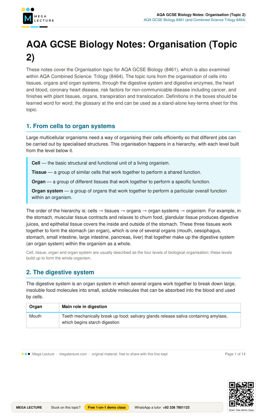
These notes cover the Organisation topic for AQA GCSE Biology (8461), which is also examined within AQA Combined Science: Trilogy (8464). The topic runs from the organisation of cells into tissues, organs and organ systems, through the digestive system and digestive enzymes, the heart and blood, coronary heart disease, risk factors for non-communicable disease including cancer, and finishes with plant tissues, organs, transpiration and translocation. Definitions in the boxes should be learned word for word; the glossary at the end can be used as a stand-alone key-terms sheet for this topic.
1. From cells to organ systems
Large multicellular organisms need a way of organising their cells efficiently so that different jobs can be carried out by specialised structures. This organisation happens in a hierarchy, with each level built from the level below it.
Cell — the basic structural and functional unit of a living organism.
Tissue — a group of similar cells that work together to perform a shared function.
Organ — a group of different tissues that work together to perform a specific function.
Organ system — a group of organs that work together to perform a particular overall function within an organism.
The order of the hierarchy is: cells → tissues → organs → organ systems → organism. For example, in the stomach, muscular tissue contracts and relaxes to churn food, glandular tissue produces digestive juices, and epithelial tissue covers the inside and outside of the stomach. These three tissues work together to form the stomach (an organ), which is one of several organs (mouth, oesophagus, stomach, small intestine, large intestine, pancreas, liver) that together make up the digestive system (an organ system) within the organism as a whole.
Cell, tissue, organ and organ system are usually described as the four levels of biological organisation; these levels build up to form the whole organism.
2. The digestive system
The digestive system is an organ system in which several organs work together to break down large, insoluble food molecules into small, soluble molecules that can be absorbed into the blood and used by cells.
| Organ | Main role in digestion |
|---|---|
| Mouth | Teeth mechanically break up food; salivary glands release saliva containing amylase, which begins starch digestion |
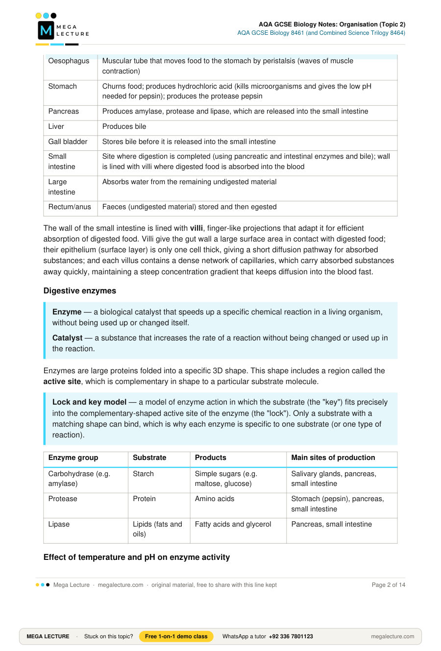
| Oesophagus | Muscular tube that moves food to the stomach by peristalsis (waves of muscle contraction) |
| Stomach | Churns food; produces hydrochloric acid (kills microorganisms and gives the low pH needed for pepsin); produces the protease pepsin |
| Pancreas | Produces amylase, protease and lipase, which are released into the small intestine |
| Liver | Produces bile |
| Gall bladder | Stores bile before it is released into the small intestine |
| Small intestine | Site where digestion is completed (using pancreatic and intestinal enzymes and bile); wall is lined with villi where digested food is absorbed into the blood |
| Large intestine | Absorbs water from the remaining undigested material |
| Rectum/anus | Faeces (undigested material) stored and then egested |
The wall of the small intestine is lined with villi, finger-like projections that adapt it for efficient absorption of digested food. Villi give the gut wall a large surface area in contact with digested food; their epithelium (surface layer) is only one cell thick, giving a short diffusion pathway for absorbed substances; and each villus contains a dense network of capillaries, which carry absorbed substances away quickly, maintaining a steep concentration gradient that keeps diffusion into the blood fast.
Digestive enzymes
Enzyme — a biological catalyst that speeds up a specific chemical reaction in a living organism, without being used up or changed itself.
Catalyst — a substance that increases the rate of a reaction without being changed or used up in the reaction.
Enzymes are large proteins folded into a specific 3D shape. This shape includes a region called the active site, which is complementary in shape to a particular substrate molecule.
Lock and key model — a model of enzyme action in which the substrate (the "key") fits precisely into the complementary-shaped active site of the enzyme (the "lock"). Only a substrate with a matching shape can bind, which is why each enzyme is specific to one substrate (or one type of reaction).
| Enzyme group | Substrate | Products | Main sites of production |
|---|---|---|---|
| Carbohydrase (e.g. amylase) | Starch | Simple sugars (e.g. maltose, glucose) | Salivary glands, pancreas, small intestine |
| Protease | Protein | Amino acids | Stomach (pepsin), pancreas, small intestine |
| Lipase | Lipids (fats and oils) | Fatty acids and glycerol | Pancreas, small intestine |
Effect of temperature and pH on enzyme activity
Every enzyme has an optimum temperature and an optimum pH at which it works fastest. As temperature rises towards the optimum, particles move faster and collide more often and with more energy, so the rate of reaction increases. Above the optimum temperature, the extra energy causes bonds holding the enzyme's 3D shape together to break; the active site changes shape and no longer fits the substrate. The enzyme is said to be denatured and can no longer catalyse the reaction, even though the temperature drops again afterwards. A pH that is too high or too low (away from the optimum) has a similar denaturing effect on the active site's shape. This is why pepsin (in the acidic stomach, optimum pH around 2) and amylase in the small intestine (optimum around pH 7–8, since the small intestine is neutral/slightly alkaline) work best in very different conditions.
Exact optimum pH values are not required knowledge for the exam — the key idea examined is that different enzymes have different optimum pH values, suited to where in the body they work.
Worked example. A student adds amylase to a starch solution and tests a drop of the mixture with iodine solution every 30 seconds. At 35 °C the mixture stops turning blue-black (i.e. all the starch has been digested) after 40 seconds. Calculate the rate of reaction in this experiment.
Rate = 1 ÷ time taken
Rate = 1 ÷ 40 = 0.025 s−1
The shorter the time taken for starch to disappear, the faster the rate of reaction, so rate is often calculated as 1 ÷ time in this type of enzyme practical.
Required practical: effect of pH on amylase activity. Amylase solution, starch solution and a buffer solution of known pH are mixed together at a fixed temperature. Every 30 seconds, a drop of the mixture is placed onto a spotting tile containing iodine solution. The end-point is reached when a spot no longer turns blue-black, showing that all the starch has been digested. The investigation is repeated using buffer solutions of different pH values, keeping everything else the same, and the time taken to reach the end-point at each pH is used to calculate the rate of reaction (rate = 1 ÷ time).
Independent variable: pH (set using different buffer solutions).
Dependent variable: time taken to reach the iodine end-point (used to calculate rate).
Control variables: concentration and volume of amylase solution; concentration and volume of starch solution; temperature.
Bile
Bile — an alkaline fluid produced by the liver, stored in the gall bladder and released into the small intestine, which neutralises stomach acid and emulsifies fats.
Emulsification — breaking large droplets of fat into many smaller droplets, greatly increasing their surface area.
Bile has two functions. First, it neutralises the hydrochloric acid brought into the small intestine from the stomach, giving an alkaline pH that is optimum for amylase, protease and lipase in the small intestine. Second, bile emulsifies fats: it breaks large fat droplets into many smaller droplets. This greatly increases the surface area of fat exposed to lipase, so fats are digested faster.
Food tests
| Food group tested | Reagent/method | Positive result |
|---|---|---|
| Starch | Add a few drops of iodine solution to the food sample | Colour changes from browny-orange to blue-black |
| Sugars (reducing sugars) | Add Benedict's solution and heat in a water bath | Colour changes from blue to green, yellow or brick-red (an orange-red precipitate forms) |
| Protein | Add sodium hydroxide solution, then a few drops of dilute copper sulfate solution (biuret test) | Colour changes from blue to purple/lilac |
| Lipids (fats) | Shake the sample with ethanol, then pour the mixture into water | A cloudy white emulsion forms |
3. The heart and blood vessels
Humans have a double circulatory system: blood passes through the heart twice for each complete circuit of the body. One circuit (the pulmonary circuit) carries deoxygenated blood from the heart to the lungs and returns oxygenated blood to the heart; the other circuit (the systemic circuit) carries oxygenated blood from the heart to the rest of the body and returns deoxygenated blood to the heart. This keeps oxygenated and deoxygenated blood separate and allows blood to be re-pressurised between the two circuits, so oxygen can be delivered to the body efficiently.
The heart has four chambers. The right atrium receives deoxygenated blood from the body through the vena cava and passes it to the right ventricle, which pumps it to the lungs through the pulmonary artery. Oxygenated blood returns from the lungs through the pulmonary vein into the left atrium and passes into the left ventricle, which pumps it to the body through the aorta. The left ventricle has a thicker muscular wall than the right ventricle because it must pump blood at higher pressure all the way around the body, whereas the right ventricle only pumps blood to the nearby lungs. A muscular wall called the septum separates the two sides of the heart and stops oxygenated and deoxygenated blood mixing.
Valves (the tricuspid valve on the right, the bicuspid/mitral valve on the left, and semilunar valves in the aorta and pulmonary artery) prevent blood flowing backwards as the heart contracts. The heart muscle itself is supplied with oxygenated blood by the coronary arteries, which branch off the aorta. The natural resting heart rate is controlled by a small group of cells in the right atrium called the pacemaker (sinoatrial node); an artificial pacemaker is a small electrical device that can be implanted to correct irregularities in heart rhythm.
Worked example. Cardiac output is the volume of blood pumped by the heart per minute.
Cardiac output (cm3/min) = heart rate (beats/min) × stroke volume (cm3/beat)
A person has a resting heart rate of 72 beats per minute and a stroke volume of 70 cm3. Calculate their cardiac output.
Cardiac output = 72 × 70 = 5040 cm3/min
Blood vessels
| Vessel | Direction of flow | Structure | Function |
|---|---|---|---|
| Artery | Away from the heart | Thick wall with muscle and elastic fibres; narrow lumen; no valves | Carries blood at high pressure; the thick elastic wall withstands and evens out the pressure surges produced by the heart |
| Vein | Towards the heart | Thinner wall than an artery; wide lumen; contains valves | Carries blood at low pressure; valves stop blood flowing backwards |
| Capillary | Links arteries to veins, running through tissues | Wall is a single cell thick; narrow lumen (about the width of a red blood cell) | Permeable walls and a large surface area allow efficient exchange of substances (oxygen, carbon dioxide, glucose, etc.) with body cells |
Note: arteries and veins are classified by the direction of blood flow (away from or towards the heart), not by whether the blood is oxygenated. The pulmonary artery carries deoxygenated blood from the heart to the lungs, and the pulmonary vein carries oxygenated blood from the lungs to the heart — the opposite of most other arteries and veins in the body.
Blood components
| Component | Key features | Function |
|---|---|---|
| Red blood cell (erythrocyte) | Biconcave disc shape; no nucleus; contains haemoglobin | Carries oxygen; haemoglobin binds oxygen in the lungs to form oxyhaemoglobin and releases it in respiring tissues. The biconcave shape gives a large surface area for gas exchange and the lack of a nucleus leaves more room for haemoglobin |
| White blood cell (leucocyte) | Has a nucleus; several types, including phagocytes and lymphocytes | Part of the immune system; phagocytes engulf and digest pathogens; lymphocytes produce antibodies (which target pathogens) and antitoxins (which neutralise toxins) |
| Platelet | Small cell fragment; no nucleus | Helps blood clot at a wound, sealing the wound and preventing blood loss and the entry of pathogens |
| Plasma | Pale yellow liquid | Transports blood cells; carries dissolved substances including glucose, amino acids, carbon dioxide, urea, hormones and antibodies |
4. Coronary heart disease and its treatment
The coronary arteries supply the heart muscle itself with oxygenated blood. In coronary heart disease, layers of fatty material build up inside the coronary arteries, narrowing them. This reduces the flow of blood through the coronary arteries, so less oxygen reaches the heart muscle. This can cause chest pain (angina) and, if an artery becomes completely blocked, a heart attack, because part of the heart muscle is starved of oxygen and stops working.
| Treatment | How it works | Advantages | Disadvantages |
|---|---|---|---|
| Stent | A small mesh tube inserted into a narrowed coronary artery to hold it open and keep blood flowing | Restores blood flow quickly; less invasive and shorter recovery than heart surgery | Surgical risks (bleeding, infection); does not stop fatty deposits building up elsewhere; may need repeating |
| Statin | A drug taken regularly that reduces the amount of cholesterol carried in the blood, slowing further build-up of fatty deposits in the arteries | Non-invasive (tablet); reduces the risk of future heart attacks and strokes | Can cause side effects (e.g. muscle pain, liver problems); must be taken long term; does not remove existing blockages |
| Artificial heart valve | A faulty valve (which lets blood leak backwards) is replaced with a mechanical valve or a biological valve made from animal tissue | Restores normal one-way blood flow through the heart | Requires open-heart surgery; mechanical valves usually require the patient to take blood-thinning (anticoagulant) drugs for life, while biological valves wear out and may need replacing |
| Heart transplant | A diseased heart is replaced with a healthy donor heart | Can save the life of a patient whose heart is failing and cannot be treated another way | Shortage of donor hearts; major surgery with recovery risks; risk of the body rejecting the organ, so the patient must take immunosuppressant drugs, which increase the risk of infection |
5. Non-communicable diseases and risk factors, including cancer
Non-communicable disease — a disease that cannot be transmitted between individuals or organisms, for example coronary heart disease, cancer or type 2 diabetes.
Risk factor — something (a substance, a characteristic of an organism, or a feature of a lifestyle) that is linked to an increased chance of developing a disease.
Many non-communicable diseases are linked to aspects of diet, lifestyle and genetics. For example, a diet high in saturated fat is linked to higher blood cholesterol and an increased risk of coronary heart disease; a diet high in sugar combined with low activity levels is linked to obesity and an increased risk of type 2 diabetes; smoking introduces tar (linked to lung disease and cancer), carbon monoxide (which reduces the blood's oxygen-carrying capacity) and nicotine (which raises heart rate and blood pressure, increasing the risk of coronary heart disease); and excessive alcohol consumption is linked to liver disease. It is important to distinguish a correlation (a relationship shown by data between a factor and a disease) from a proven cause: further evidence, such as a plausible biological mechanism, is needed before a correlation can be treated as causal.
Obesity is itself a risk factor for several non-communicable diseases, including type 2 diabetes and cardiovascular disease. It is often assessed using Body Mass Index (BMI), calculated as BMI = mass (kg) ÷ [height (m)]2. A BMI of 18.5–24.9 is classed as a healthy weight, 25–29.9 as overweight, and 30 or above as obese. Waist:hip ratio (waist circumference ÷ hip circumference) is sometimes used instead of BMI, since it better reflects the distribution of body fat and can be a stronger indicator of the risk of conditions such as type 2 diabetes and cardiovascular disease.
Worked example. Calculate the BMI of a person with a mass of 70 kg and a height of 1.75 m.
BMI = mass (kg) ÷ [height (m)]2
BMI = 70 ÷ 1.752 = 70 ÷ 3.0625 = 22.9 (3 s.f.)
This BMI falls within the healthy weight range (18.5–24.9).
Non-communicable diseases also carry a human and financial cost. For the individual, this can include a reduced quality of life, lost earnings if they are unable to work, and the cost of their own treatment or ongoing care. For the local community and national health service, costs include the price of drugs and treatment, hospital beds, and staff time that could otherwise be used for other patients. Nationally and globally, non-communicable diseases reduce productivity through days lost from work or school, and the growing number of people living with these conditions places an increasing financial burden on health and social care systems.
Cancer
Tumour — a mass of abnormal cells that results from uncontrolled cell division/growth.
Benign tumour — a tumour that grows in one place, usually within a membrane, and does not invade or spread to other tissues.
Malignant tumour — a cancerous tumour that invades neighbouring tissue and can spread, via the blood or lymph, to form secondary tumours in other parts of the body (metastasis).
Cancer is caused by changes in cells that lead to uncontrolled growth and division. Risk factors that increase the chance of cancer developing include genetic factors (some gene variants are inherited and increase risk, for example of breast or bowel cancer), carcinogens (cancer-causing substances or agents, such as chemicals in tobacco smoke, ultraviolet radiation from sunlight, and ionising radiation), certain viral infections, and lifestyle factors such as obesity and diet. There are many different types of cancer, which can affect almost any tissue or organ; the specific risk factors, typical age of onset and outlook vary considerably between different types of cancer. For example, lung cancer is strongly linked to carcinogens in tobacco smoke, skin cancer is linked to exposure to ultraviolet radiation, cervical cancer is linked to certain strains of HPV (human papillomavirus) infection, and some breast and bowel cancers are linked to inherited gene variants.
6. Plant tissues and organs
Just as in animals, plant cells are organised into tissues, organs and organ systems.
| Plant tissue | Description | Function |
|---|---|---|
| Epidermal tissue | A single layer of cells covering the plant, often coated with a waxy cuticle | Protects the plant and reduces water loss |
| Palisade mesophyll | Tall, closely packed cells containing many chloroplasts, found near the upper surface of a leaf | The main site of photosynthesis, positioned to absorb maximum light |
| Spongy mesophyll | Irregularly shaped cells with large air spaces between them, found below the palisade layer | Air spaces allow carbon dioxide and oxygen to diffuse to and from the palisade cells and out through the stomata |
| Xylem | Tube-like tissue made of dead, hollow cells strengthened with lignin | Transports water and dissolved mineral ions from the roots to the leaves (one direction only) |
| Phloem | Tissue made of living cells (sieve tube elements and companion cells) | Transports dissolved sugars and amino acids around the plant (translocation), in both directions |
| Meristem tissue | Found at the growing tips of roots and shoots; cells can divide and differentiate into any type of plant cell | Enables plant growth |
The leaf is a plant organ built from several of these tissues working together. From top to bottom, a typical leaf is organised as: waxy cuticle, upper epidermis, palisade mesophyll, spongy mesophyll, lower epidermis (containing pairs of guard cells surrounding pores called stomata), with vascular bundles of xylem and phloem running through the leaf. Other plant organs include the stem, the root and the flower; together they form the plant's organ systems, such as the transport (vascular) system made of xylem and phloem.
7. Transpiration and translocation
Transpiration — the loss of water vapour from a plant's surface (mainly through stomata in the leaves) by evaporation and diffusion.
Translocation — the movement of dissolved sugars (mainly sucrose) and amino acids through phloem tissue, from sources (where they are made or stored) to sinks (where they are used or stored), in either direction.
Water is drawn up from the roots, through the xylem, and out through the stomata of the leaves as part of a continuous flow called the transpiration stream. Water enters root hair cells by osmosis, moves across the root into the xylem, travels up the stem in the xylem, and evaporates from the surface of mesophyll cells inside the leaf before diffusing out through open stomata. This flow replaces the water lost in transpiration and also carries dissolved mineral ions upward through the plant.
Translocation moves the sugars made in photosynthesis (mainly in mature leaves, which act as the "source") to parts of the plant that need sugars for respiration, growth or storage, such as roots, buds and developing fruit or seeds (which act as "sinks"). Unlike the one-way transpiration stream in xylem, translocation in phloem can occur in more than one direction around the plant, depending on where sources and sinks are located, and requires the plant to use energy.
Factors affecting the rate of transpiration
| Factor | Effect on rate of transpiration | Reason |
|---|---|---|
| Light intensity | Higher light intensity increases the rate | Stomata open wider in the light to let in carbon dioxide for photosynthesis, so more water vapour can escape; in the dark, stomata close and transpiration falls almost to zero |
| Temperature | Higher temperature increases the rate | Water particles have more kinetic energy, so evaporation from cells inside the leaf and diffusion of water vapour out of the stomata both speed up |
| Humidity | Higher humidity decreases the rate | A smaller difference in water vapour concentration between the air inside the leaf and the air outside reduces the diffusion gradient, so less water vapour diffuses out |
| Air movement (wind) | Greater air movement increases the rate | Moving air carries away water vapour as soon as it diffuses out of the stomata, maintaining a steep concentration gradient that keeps diffusion (and therefore transpiration) fast |
Worked example. A potometer is used to estimate the rate of transpiration by measuring the rate of water uptake by a shoot. An air bubble in the capillary tube of a potometer moves 24 mm in 2 minutes. The capillary tube has a radius of 0.5 mm. Calculate the rate of water uptake.
Cross-sectional area of tube = π × r2 = π × 0.52 = 0.7854 mm2 (0.79 mm2 to 2 s.f.)
Volume of water taken up = cross-sectional area × distance moved = 0.7854 × 24 = 18.8 mm3 (using the unrounded area to avoid a rounding error)
Rate of water uptake = volume ÷ time = 18.8 ÷ 2 = 9.4 mm3/min
Carry unrounded figures through a multi-step calculation and only round the final answer, to avoid introducing rounding errors.
A potometer measures water uptake by the shoot, which is used as an estimate of water loss by transpiration, since most water taken up by a leafy shoot is lost through transpiration rather than used in other processes.
Key terms
| Term | Definition |
|---|---|
| Cell | The basic structural and functional unit of a living organism |
| Tissue | A group of similar cells that work together to carry out a shared function |
| Organ | A group of different tissues that work together to perform a specific function |
| Organ system | A group of organs that work together to perform a particular overall function |
| Muscular tissue | Tissue that contracts (shortens) to bring about movement |
| Glandular tissue | Tissue that produces and secretes substances such as enzymes and hormones |
| Epithelial tissue | Tissue that covers body surfaces, including the inside and outside of organs |
| Enzyme | A biological catalyst; a protein that speeds up a specific reaction without being used up |
| Catalyst | A substance that increases the rate of a reaction without being used up or changed |
| Substrate | The molecule that an enzyme acts upon in a reaction |
| Active site | The region of an enzyme with a shape complementary to its substrate, where the reaction takes place |
| Lock and key model | A model of enzyme action in which a substrate (key) binds only to an enzyme's complementary-shaped active site (lock) |
| Denatured | Describes an enzyme whose active site has changed shape (due to high temperature or extreme pH) so it can no longer bind its substrate |
| Optimum temperature | The temperature at which an enzyme works fastest |
| Optimum pH | The pH at which an enzyme works fastest |
| Carbohydrase | An enzyme that breaks down carbohydrates (e.g. starch) into simple sugars |
| Amylase | A carbohydrase enzyme that breaks down starch into sugars |
| Protease | An enzyme that breaks down proteins into amino acids |
| Lipase | An enzyme that breaks down lipids into fatty acids and glycerol |
| Bile | An alkaline fluid, made in the liver and stored in the gall bladder, that neutralises stomach acid and emulsifies fats |
| Emulsification | The breaking up of large fat droplets into many smaller droplets, increasing their surface area |
| Peristalsis | Waves of muscular contraction that push food along the gut |
| Villus (plural: villi) | A finger-like projection of the small intestine wall that increases the surface area for absorption |
| Absorption | The movement of digested food molecules from the gut into the blood |
| Egestion | The removal of undigested food (faeces) from the body |
| Reducing sugar | A sugar that gives a positive (colour-change) result with Benedict's solution on heating |
| Biuret test | A chemical test for protein, giving a purple/lilac colour with sodium hydroxide and copper sulfate solution |
| Double circulatory system | A circulatory system in which blood passes through the heart twice for each full circuit of the body: once to the lungs and once to the rest of the body |
| Atrium | An upper chamber of the heart that receives blood returning to the heart |
| Ventricle | A lower chamber of the heart that pumps blood out of the heart |
| Septum | The muscular wall separating the left and right sides of the heart, preventing blood mixing |
| Valve | A structure in the heart or veins that prevents blood flowing backwards |
| Pacemaker | A group of cells in the right atrium that controls the natural resting heart rate (or an artificial device that performs this function) |
| Aorta | The main artery carrying oxygenated blood from the left ventricle to the body |
| Vena cava | The main vein carrying deoxygenated blood from the body into the right atrium |
| Pulmonary artery | The blood vessel carrying deoxygenated blood from the right ventricle to the lungs |
| Pulmonary vein | The blood vessel carrying oxygenated blood from the lungs to the left atrium |
| Coronary artery | A blood vessel that supplies the heart muscle itself with oxygenated blood |
| Cardiac output | The volume of blood pumped by the heart per minute (heart rate × stroke volume) |
| Stroke volume | The volume of blood pumped out of the heart with each beat |
| Artery | A blood vessel with a thick, muscular, elastic wall that carries blood away from the heart at high pressure |
| Vein | A blood vessel with a wide lumen and valves that carries blood towards the heart at low pressure |
| Capillary | A microscopic blood vessel with a wall one cell thick, allowing exchange of substances with tissues |
| Red blood cell (erythrocyte) | A biconcave, nucleus-free blood cell containing haemoglobin, which carries oxygen |
| Haemoglobin | The red pigment in red blood cells that binds oxygen to form oxyhaemoglobin |
| White blood cell (leucocyte) | A blood cell with a nucleus that is part of the immune system, defending against pathogens |
| Phagocyte | A type of white blood cell that engulfs and digests pathogens |
| Lymphocyte | A type of white blood cell that produces antibodies and antitoxins |
| Antibody | A protein produced by lymphocytes that targets a specific pathogen |
| Antitoxin | A substance produced by lymphocytes that neutralises toxins produced by pathogens |
| Platelet | A small cell fragment involved in blood clotting at wound sites |
| Plasma | The pale yellow liquid part of blood that transports blood cells and dissolved substances |
| Coronary heart disease | A disease in which fatty deposits narrow the coronary arteries, reducing blood flow to the heart muscle |
| Stent | A mesh tube inserted into a narrowed artery to hold it open |
| Statin | A drug that reduces blood cholesterol level and slows the build-up of fatty deposits in arteries |
| Non-communicable disease | A disease that cannot be passed between individuals |
| Risk factor | A factor linked to an increased chance of developing a disease |
| Body Mass Index (BMI) | A measure of body mass relative to height, calculated as mass (kg) ÷ [height (m)]2, used to assess whether a person is a healthy weight, overweight or obese |
| Waist:hip ratio | Waist circumference divided by hip circumference; sometimes used instead of BMI to assess the risk of conditions such as type 2 diabetes and cardiovascular disease |
| Correlation | A relationship between two factors shown by data, which may or may not be causal |
| Tumour | A mass of abnormal cells resulting from uncontrolled cell division |
| Benign tumour | A tumour that grows in one place and does not spread to other tissues |
| Malignant tumour | A cancerous tumour that invades and spreads to other tissues, forming secondary tumours |
| Metastasis | The spread of cancer cells from a primary tumour to form secondary tumours elsewhere in the body |
| Carcinogen | A substance or agent that can cause cancer |
| Epidermal tissue | The outer covering tissue of a plant, often coated with a waxy cuticle |
| Palisade mesophyll | Tightly packed, chloroplast-rich leaf tissue that is the main site of photosynthesis |
| Spongy mesophyll | Leaf tissue with air spaces that allows diffusion of gases within the leaf |
| Xylem | Plant tissue made of dead, lignified cells that transports water and mineral ions from roots to leaves |
| Phloem | Plant tissue made of living cells that transports dissolved sugars and amino acids around the plant |
| Meristem tissue | Undifferentiated plant tissue at growing tips, able to divide and form any type of plant cell |
| Stomata | Pores, usually in the lower epidermis of a leaf, through which gases and water vapour move |
| Guard cells | Pairs of cells that change shape to open or close a stoma |
| Transpiration | The loss of water vapour from a plant's surface, mainly through the stomata |
| Transpiration stream | The continuous one-way flow of water from roots to leaves through the xylem |
| Translocation | The movement of dissolved sugars and amino acids through phloem, from sources to sinks |
| Potometer | Apparatus used to estimate the rate of transpiration by measuring the rate of water uptake by a shoot |
Quick check
- Name the four levels of organisation in a multicellular organism, from smallest to largest. [4]
- State the product of the digestion of protein by protease. [1]
- Give the organ that produces bile and the organ that stores it. [2]
- Describe two ways in which the villi of the small intestine are adapted to maximise the rate of absorption of digested food. [4]
- A student adds iodine solution to a food sample and it stays browny-orange. State what this shows about the sample. [1]
- Describe how bile helps in the digestion of fats. [3]
- Explain why an enzyme stops working if the temperature rises well above its optimum. [3]
- Compare the structure of arteries and veins, and explain how the structure of each is related to its function. [4]
- State which blood vessel carries deoxygenated blood away from the heart, and explain why this appears to break the "usual" rule linking arteries to oxygenated blood. [2]
- Calculate the cardiac output of a person with a heart rate of 65 beats per minute and a stroke volume of 80 cm3. Give the unit. [2]
- Explain how a stent and a statin each reduce the risk of a heart attack in a patient with coronary heart disease. [4]
- Calculate the BMI of a person with a mass of 84 kg and a height of 1.8 m, and state whether this places them in the healthy, overweight or obese range. [3]
- Explain two ways in which a rise in obesity in a population could increase costs to a national health service. [4]
- A tumour is found to be contained within a membrane and has not spread to other tissues. State whether this is a benign or a malignant tumour, and give one further difference between the two types of tumour. [2]
- Describe the structure of xylem tissue and explain how this structure is related to its function. [3]
- Explain why increasing air movement around a leafy shoot increases its rate of transpiration. [3]
- Evaluate the use of an artificial heart valve compared with a donor heart transplant to treat a patient with severe heart valve disease. [6]
1. Cells → tissues → organs → organ systems.
2. Amino acids.
3. Produced by the liver; stored in the gall bladder.
4. Any two of: villi have a large surface area, increasing the area available for absorption, so more digested food can be absorbed at once; the villus wall/epithelium is one cell thick, giving a short diffusion pathway, so substances diffuse into the blood more quickly; villi have a dense network of capillaries (a good blood supply), which carries absorbed substances away quickly and maintains a steep concentration gradient for diffusion.
5. No starch is present in the sample.
6. Bile is alkaline and neutralises the acid brought into the small intestine from the stomach, giving a suitable (alkaline) pH for enzymes such as lipase; bile also emulsifies fats, breaking large fat droplets into smaller droplets, which increases the surface area of fat, so lipase can digest it faster.
7. At high temperatures the enzyme's molecule vibrates more, breaking the bonds that hold its 3D shape together; the active site changes shape; the substrate can no longer bind (fit) the active site, so the enzyme is denatured and can no longer catalyse the reaction.
8. Arteries have thick, muscular, elastic walls and a narrow lumen, and no valves; this withstands and evens out the high pressure of blood pumped from the heart. Veins have thinner walls, a wider lumen, and contain valves; this suits the lower pressure blood flow and the valves stop blood flowing backwards on its way to the heart.
9. The pulmonary artery. Arteries and veins are classified by the direction of blood flow (away from or towards the heart), not by the oxygen content of the blood — the pulmonary artery carries deoxygenated blood from the heart to the lungs, which is the opposite of the oxygen content usually associated with arteries.
10. Cardiac output = 65 × 80 = 5200 cm3/min.
11. A stent is inserted into a narrowed coronary artery and holds it open, restoring blood flow to the heart muscle immediately. A statin is a drug that lowers blood cholesterol level, which slows down the rate at which fatty deposits build up in the coronary arteries, reducing the risk of them narrowing further in future.
12. BMI = 84 ÷ 1.82 = 84 ÷ 3.24 = 25.9 (3 s.f.); this places them in the overweight range (25–29.9).
13. Any two of, each with a linked explanation: more people develop obesity-related conditions such as type 2 diabetes, cardiovascular disease or some cancers, increasing the cost of drugs, hospital beds and staff time needed to treat them; more people may be too unwell to work, reducing productivity/tax revenue and increasing the cost of benefits or long-term care; increased demand for treatments such as weight-loss surgery adds further costs to the health service.
14. Benign; a malignant tumour invades neighbouring tissue and can spread to other parts of the body (via the blood or lymph) to form secondary tumours, whereas a benign tumour does not spread to other tissues.
15. Xylem is made of dead, hollow cells joined end to end to form long, continuous tubes; the cell walls are strengthened/thickened with lignin. The hollow tube structure allows water (and dissolved mineral ions) to flow through easily, and the lignin gives the tissue strength to withstand the pressure changes involved and to support the plant.
16. Moving air removes water vapour molecules from the air just outside the stomata as they diffuse out; this keeps the concentration of water vapour outside the leaf lower than inside, maintaining a steep diffusion gradient, so water vapour continues to diffuse out of the stomata quickly, increasing the transpiration rate.
17. Both treatments restore normal blood flow through the heart, but they differ in scale and risk. An artificial valve replaces only the faulty valve, is more widely available than a donor heart, and involves a single operation, but mechanical valves usually require the patient to take anticoagulant drugs for life (risk of bleeding) while biological valves wear out and may eventually need replacing; a heart transplant replaces the whole heart and can be the only option when the heart muscle itself is failing, but donor hearts are in short supply, the surgery carries greater risk, and the patient must take immunosuppressant drugs for life, which increases their risk of infection. Overall, valve replacement is usually preferred when only the valve is faulty, since it is less invasive and does not depend on a compatible donor organ, while transplant is reserved for cases where the whole heart is failing.
