
IB Diploma · Biology · SL / HL · Theme B: Form and Function
B1.2 Proteins
Opens inside this page. Prefer your own PDF app? Open the PDF.
Full text of B1.2 Proteins
IB DP BIOLOGY Theme B: Form and Function B1.2 Proteins
Revision Notes · Standard and Higher Level Fahad H. Ahmad
+92 323 509 4443 | Megalecture.com
Original notes prepared for the IB Diploma Programme Biology course (first assessment 2025)
What the syllabus requires
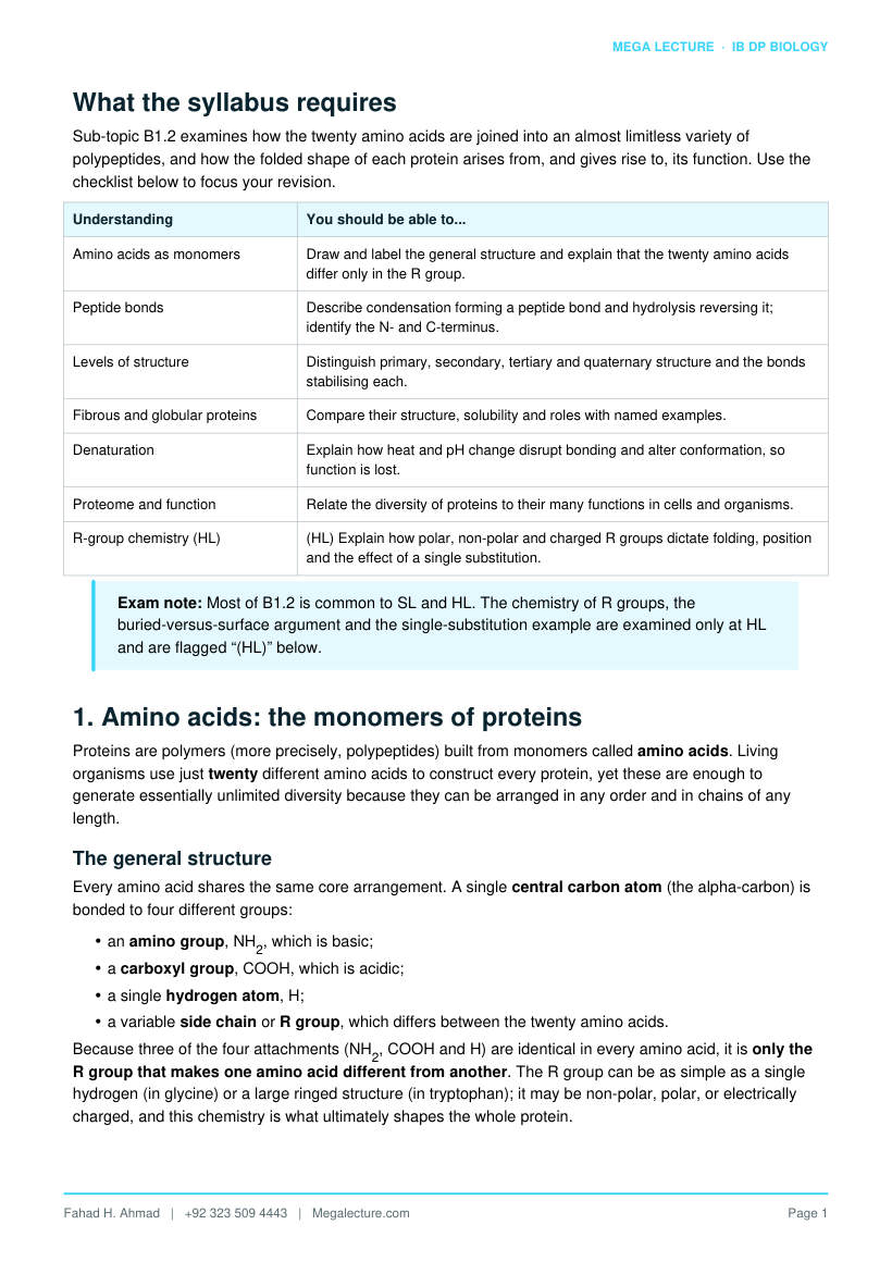
Sub-topic B1.2 examines how the twenty amino acids are joined into an almost limitless variety of polypeptides, and how the folded shape of each protein arises from, and gives rise to, its function. Use the checklist below to focus your revision.
Understanding You should be able to...
Amino acids as monomers Draw and label the general structure and explain that the twenty amino acids differ only in the R group.
Peptide bonds Describe condensation forming a peptide bond and hydrolysis reversing it; identify the N- and C-terminus.
Levels of structure Distinguish primary, secondary, tertiary and quaternary structure and the bonds stabilising each.
Fibrous and globular proteins Compare their structure, solubility and roles with named examples.
Denaturation Explain how heat and pH change disrupt bonding and alter conformation, so function is lost.
Proteome and function Relate the diversity of proteins to their many functions in cells and organisms.
R-group chemistry (HL) (HL) Explain how polar, non-polar and charged R groups dictate folding, position and the effect of a single substitution.
Exam note: Most of B1.2 is common to SL and HL. The chemistry of R groups, the buried-versus-surface argument and the single-substitution example are examined only at HL and are flagged “(HL)” below.
1. Amino acids: the monomers of proteins
Proteins are polymers (more precisely, polypeptides) built from monomers called amino acids. Living organisms use just twenty different amino acids to construct every protein, yet these are enough to generate essentially unlimited diversity because they can be arranged in any order and in chains of any length.
The general structure
Every amino acid shares the same core arrangement. A single central carbon atom (the alpha-carbon) is bonded to four different groups:
- an amino group, NH2, which is basic;
- a carboxyl group, COOH, which is acidic;
- a single hydrogen atom, H;
- a variable side chain or R group, which differs between the twenty amino acids.
Because three of the four attachments (NH2, COOH and H) are identical in every amino acid, it is only the R group that makes one amino acid different from another. The R group can be as simple as a single hydrogen (in glycine) or a large ringed structure (in tryptophan); it may be non-polar, polar, or electrically charged, and this chemistry is what ultimately shapes the whole protein.
Draw it correctly: In an exam diagram place the central carbon in the middle, with NH2 on the left, COOH on the right, H below and R above. Label all four groups. Marks are commonly lost for omitting the hydrogen or mislabelling the amino and carboxyl groups.
Essential and non-essential amino acids
Of the twenty amino acids, some can be made by the human body from other molecules (non-essential amino acids) while others cannot be synthesised and must be obtained ready-made from the diet (essential amino acids). This explains why a balanced supply of dietary protein matters: a diet short of one essential amino acid limits the proteins the body can build.
Charge at cell pH
At the pH inside cells an amino acid does not carry neutral NH2 and COOH groups. The carboxyl group tends to lose a hydrogen ion (becoming negatively charged) and the amino group tends to gain one (becoming positively charged), so the molecule carries a positive and a negative charge at the same time. This dual-charged form explains why R-group charge is so sensitive to pH, a point developed in section 10.
2. Peptide bonds: joining the monomers
Amino acids are linked by condensation reactions. The carboxyl group (COOH) of one amino acid reacts with the amino group (NH2) of the next; a molecule of water is removed, and a new covalent bond called a peptide bond forms between the carbon and the nitrogen.
amino acid + amino acid → dipeptide + water
Two amino acids joined this way form a dipeptide; a chain of many amino acids joined by many peptide bonds is a polypeptide. A functional protein consists of one or more polypeptides. Each additional amino acid added to the chain releases one further molecule of water, so a polypeptide of n amino acids is formed with the loss of n − 1 water molecules.
Hydrolysis: the reverse
Peptide bonds are broken by hydrolysis, the reverse reaction, in which a water molecule is added across the bond to separate the two amino acids. This is how digestive enzymes (proteases such as pepsin and trypsin) break dietary protein into amino acids for absorption, and how cells recycle their own proteins.
Directionality: N-terminus and C-terminus
A polypeptide has direction. At one end there is a free amino group (the N-terminus) and at the other a free carboxyl group (the C-terminus). By convention the sequence is written and the chain is synthesised on the ribosome starting from the N-terminus. This directionality matters because the same amino acids in reverse order give a different protein.
Common misconception: Peptide bonds form by condensation (removing water), not by hydrolysis. Confusing the two, or writing that condensation uses up water, is one of the most frequent errors in this topic.
Figure 1. Condensation of two amino acids. The −OH of one carboxyl group and an H− of the next amino group are removed as 1 water molecule (n−1 per chain), forming a peptide bond (CO−NH). Hydrolysis reverses the reaction.
3. Primary structure
The primary structure is the specific sequence of amino acids in a polypeptide, held together by peptide bonds. It is written from the N-terminus to the C-terminus and is the simplest, most fundamental level of protein structure.
The sequence is determined by the gene that codes for the protein: the order of bases in DNA is transcribed into mRNA and then translated, three bases (a codon) at a time, into a precise order of amino acids. A change in the DNA sequence (a mutation) can therefore change the primary structure.
Why primary structure matters
The primary structure is decisive because it determines every higher level of structure. The identity and order of the R groups fix where the chain will coil, fold and cross-link, and therefore fix the final three-dimensional shape. Since shape determines function, the whole behaviour of a protein is ultimately encoded in its amino-acid sequence. Even a single incorrect amino acid can alter folding enough to change or destroy function.
Key principle: Sequence → shape → function. Almost every “explain” question in this topic is a variation on this chain of cause and effect.
4. Secondary structure
The secondary structure is the regular, repeating local folding of the polypeptide backbone into recognisable shapes. It is stabilised by hydrogen bonds that form between atoms of the backbone − specifically between the slightly positive hydrogen of an N−H group and the slightly negative oxygen of a C=O group further along the chain. The R groups are not involved. There are two common forms:
- the α-helix − the chain coils into a right-handed spiral, with hydrogen bonds running roughly parallel to the axis of the coil, holding each turn to the next;
- the β-pleated sheet − two or more sections of the chain lie side by side and are linked by hydrogen bonds between them, giving a folded, sheet-like arrangement.
A single polypeptide may contain regions of α-helix, regions of β-pleated sheet, and regions with no regular pattern. Although each individual hydrogen bond is weak, the large number of them makes secondary structure an important stabilising contribution to the overall shape.
Watch the wording: Secondary structure is stabilised by hydrogen bonds between the backbone (the C=O and N−H groups), not between R groups. R-group interactions belong to tertiary structure.
5. Tertiary structure
The tertiary structure is the overall three-dimensional shape of a single polypeptide, produced as the chain (already containing helices and sheets) folds back on itself. This folding is driven and stabilised by interactions between the R groups of amino acids that are brought close together in space, even if they are far apart in the sequence. Four types of interaction are involved:
Interaction How it forms Strength
Hydrogen bonds Between polar R groups, where a slightly positive H is attracted to a slightly negative O or N.
Weak (individually)
Ionic bonds Between R groups carrying opposite charges (one positive, one negative).
Moderate
Disulfide bridges A covalent S−S bond between the sulfur atoms of two cysteine R groups.
Strong (covalent)
Hydrophobic interactions Non-polar R groups cluster together, away from water, in the interior of the protein.
Weak but numerous
The balance of these interactions folds each polypeptide into one precise, stable shape. The disulfide bridge is the only covalent link among them and so is the strongest; the others are weaker but, acting together in large numbers, hold the structure firmly. This folded shape is what gives a protein such as an enzyme its specific active site.
Exam tip: If asked for the strongest stabilising bond in tertiary structure, name the disulfide bridge (covalent). If asked which interactions form between R groups, list all four.
6. Quaternary structure
Some proteins consist of more than one polypeptide chain. The quaternary structure is the way these separate subunits fit together, held by the same types of R-group interaction that stabilise tertiary structure. Only proteins with two or more polypeptides have a quaternary structure; a single-chain protein does not.
Many proteins also include a non-polypeptide component, called a prosthetic group, that is permanently bound and needed for the protein to work. A protein containing such a group is a conjugated protein.
Protein Quaternary arrangement Note
Haemoglobin Four polypeptide subunits (two α and two β chains), each carrying a haem group.
The four haem groups (each with an iron ion) bind oxygen; haem is the prosthetic group.
Collagen Three polypeptide chains wound around one another into a triple helix.
The rope-like structure gives great tensile strength in skin, tendon and bone.
Insulin Two short polypeptide chains (A and B) joined by disulfide bridges.
A small hormone; illustrates disulfide links between chains.
Haemoglobin is the standard example: its four subunits and four haem prosthetic groups together allow it to bind and transport four oxygen molecules, and the cooperative interaction between subunits makes oxygen uptake and release efficient.
Summary of the four levels
It is worth being able to place each level, its description and its stabilising bonds side by side, because “compare the levels of protein structure” is a recurring exam instruction.
Level What it is Held together by
Primary The sequence of amino acids in the chain Peptide bonds (covalent)
Secondary Local coiling/folding into α-helix or β-pleated sheet
Hydrogen bonds between backbone atoms
Tertiary Overall 3-D fold of one polypeptide H-bonds, ionic bonds, disulfide bridges, hydrophobic interactions (all between R groups)
Quaternary Assembly of two or more subunits (and prosthetic groups)
The same R-group interactions, between chains
Figure 2. The four levels of protein structure: primary (sequence) → secondary (α-helix and β-pleated sheet) → tertiary (3-D fold of one chain) → quaternary (4 levels in all; haemoglobin has 4 subunits).
7. Fibrous and globular proteins
Proteins fall into two broad shape classes that suit very different jobs. Fibrous proteins are long and thread-like and usually structural; globular proteins are rounded and compact and usually metabolically active.
Feature Fibrous proteins Globular proteins
Shape Long, narrow strands or fibres Rounded, compact, roughly spherical
Secondary/tertiary Highly regular, repetitive; little folding into a ball
Complex, irregular folding into a precise 3-D shape
Solubility in water Generally insoluble Generally soluble (hydrophilic R groups face outward)
Stability to change Tough, resistant, stable to changes in temperature and pH
More sensitive; readily denatured
Typical role Structural and support Functional and metabolic
Examples Collagen (connective tissue), keratin (hair, nails), silk
Haemoglobin, enzymes such as amylase, antibodies, insulin
The contrast follows directly from structure: the regular, cross-linked strands of a fibrous protein make it strong and insoluble and ideal for support, while the compact fold of a globular protein, with hydrophilic groups on its surface, makes it soluble and able to present a precise site for chemical activity.
8. Denaturation
Denaturation is the loss of a protein's specific three-dimensional shape, caused by breaking the weak interactions (hydrogen bonds, ionic bonds and hydrophobic interactions) that hold the tertiary and quaternary structure together. Because shape determines function, a denatured protein loses its function − an enzyme, for example, can no longer bind its substrate at the active site. Denaturation is usually irreversible.
Heat
Raising the temperature gives atoms more kinetic energy, so the chain vibrates more. Above a certain temperature these vibrations break the hydrogen and ionic bonds holding the fold in place, and the protein unravels into a different, non-functional conformation. Cooking an egg is the everyday example: the clear albumen turns white and solid as its proteins denature and no longer return to their original state.
pH change
A change in pH alters the concentration of hydrogen ions, which changes the charge on the acidic and basic R groups. This disrupts the ionic bonds (and some hydrogen bonds) between R groups, so the protein loses its shape. Each protein has an optimum pH; move far from it and the protein denatures. Adding acid to milk, which curdles the protein casein, is a familiar example.
Other denaturing agents
Heat and pH are the two the syllabus emphasises, but the same principle − disruption of the weak bonds that hold the fold − applies to other agents. Heavy-metal ions and some organic solvents and detergents can also denature proteins by interfering with ionic and hydrophobic interactions. In every case the pattern is the same: the peptide bonds survive, but the three-dimensional shape, and therefore the function, is lost.
Figure 3. Modelled relative activity against temperature and pH. Activity rises to a peak at the optimum (T ≈ 40°C, pH 7), then falls steeply as heat and extreme pH break the weak bonds and the protein denatures.
Common misconception: Denaturation does not break peptide bonds and does not change the primary structure. The sequence of amino acids is unchanged; only the folding (secondary, tertiary and quaternary structure) is disrupted.
9. Protein diversity, the proteome and function
Because polypeptides can be any length and any sequence of the twenty amino acids, the number of possible proteins is enormous. This diversity of shape underlies the huge range of jobs that proteins do. The complete set of proteins expressed by a cell or organism is called its proteome; unlike the genome it is not fixed, but changes with cell type, activity and conditions.
Function What the protein does Example
Catalysis Enzymes speed up metabolic reactions. Amylase, catalase
Structure and support Provide mechanical strength. Collagen, keratin
Transport Carry molecules within the body or across membranes. Haemoglobin; channel proteins
Hormones (signalling) Chemical messengers coordinating the body. Insulin
Defence Antibodies bind and neutralise pathogens. Immunoglobulins
Receptors Detect signals at the cell surface and inside cells. Membrane receptor proteins
Movement Motor proteins generate force and movement. Actin and myosin in muscle
The unifying idea is that each of these functions depends on a particular shape, and that shape comes from the primary structure. Diversity of sequence therefore produces diversity of shape, and diversity of shape produces diversity of function.
10. R-group chemistry and folding (HL)
(HL) At higher level you should explain why a polypeptide folds the way it does, in terms of the chemistry of its R groups. Amino acids are grouped by the nature of their side chain:
R group type Behaviour in water Typical position in a folded, water-based protein
Non-polar (hydrophobic)
Repelled by water; cluster together away from it.
Buried in the interior core of the protein
Polar (uncharged) Interact with water; form hydrogen bonds. Often on the surface, or lining internal channels
Charged (acidic or basic)
Strongly attracted to water; can form ionic bonds.
On the surface, in contact with the surrounding water
How R groups dictate folding and position
As a polypeptide folds in the watery cytoplasm, hydrophobic R groups are driven to the inside, forming a non-polar core, while hydrophilic (polar and charged) R groups are held on the outside, where they interact with water. This arrangement is a major reason globular proteins are soluble and fold into one stable shape. Within the core and at the surface, specific pairs of R groups then form the hydrogen bonds, ionic bonds and disulfide bridges that lock the tertiary structure. A membrane protein shows the reverse pattern: non-polar R groups face outward into the lipid bilayer.
pH and charge (HL)
(HL) The charge on acidic and basic R groups depends on pH. At low pH extra hydrogen ions neutralise negative groups; at high pH basic groups lose their positive charge. Changing these charges makes or breaks ionic bonds and alters folding − which is why pH affects both protein shape and, for enzymes, activity.
Effect of a single amino-acid change (HL)
(HL) Because folding depends on the exact R groups present, changing even one amino acid can change the whole protein. The classic conceptual example is sickle-cell anaemia: in the haemoglobin β-chain a single substitution replaces a charged, hydrophilic amino acid (glutamate) with a non-polar, hydrophobic one (valine). The new hydrophobic patch on the surface makes haemoglobin molecules stick together into fibres when oxygen is low, distorting red blood cells into a sickle shape. One change in the primary structure thus alters shape, behaviour and function − a direct demonstration of the sequence → shape → function principle.
HL link: This connects to B1.2 and to genetics: a point mutation in DNA changes one codon, which changes one amino acid, which can change the protein's R-group chemistry and therefore its folding and function.
11. Worked examples and exam skills
The questions below rehearse the four skills examiners test most often: identifying the level of structure and the bond involved, explaining denaturation, relating sequence to function, and interpreting a described structure.
Worked example 1 — identify the bond and structure level
In a protein, two cysteine side chains a long way apart in the sequence are joined by a strong covalent link. Name the bond and state which level of structure it stabilises.
The bond is a disulfide bridge (an S−S covalent bond between the R groups of two cysteines). Because it forms between R groups within one folded chain, it stabilises the tertiary structure (and, where it links two chains as in insulin, the quaternary structure). It is the strongest of the tertiary interactions.
Worked example 2 — explain denaturation
A student boils a solution of the enzyme catalase and finds it no longer breaks down hydrogen peroxide. Explain, in terms of bonding and shape, why activity is lost.
Heat gives the molecule more kinetic energy, so the chain vibrates and the hydrogen and ionic bonds holding the tertiary structure break. The protein loses its specific three-dimensional shape (it denatures), so the active site changes shape and can no longer bind hydrogen peroxide. Function is lost, and the change is irreversible.
Note: the peptide bonds and primary structure are unchanged − only the folding is disrupted.
Worked example 3 — relate sequence to function
Explain how a change in one DNA base can lead to a non-functional protein.
A change in a DNA base can change one codon, so a different amino acid is placed in the polypeptide, altering the primary structure. Because the primary structure determines folding, a different R group at that position can change the pattern of hydrogen, ionic, hydrophobic and disulfide interactions, giving a different tertiary shape. If the active site or binding region is altered, the protein cannot carry out its function. This is the sequence → shape → function chain.
Worked example 4 — interpret a described structure
A protein is described as consisting of four polypeptide subunits, each folded into a compact globular shape and each holding an iron-containing prosthetic group; it is soluble and transports oxygen in the blood. Identify the protein and the highest level of structure present, and justify your answer.
The protein is haemoglobin. Because it has more than one polypeptide subunit (four) held together, it shows quaternary structure − the highest level. The iron-containing group is the haem prosthetic group, and its compact, soluble, globular form suits its transport role.
Common pitfalls to avoid
- Writing that peptide bonds form by hydrolysis. They form by condensation, removing water; hydrolysis breaks them.
- Saying secondary structure is stabilised by R-group bonds. It is stabilised by hydrogen bonds between the backbone (C=O and N−H).
- Claiming denaturation breaks peptide bonds or changes the sequence. It disrupts folding only; the primary structure is unchanged.
- Giving a quaternary structure to a single-chain protein. Only proteins with two or more polypeptides have quaternary structure.
- Confusing the four tertiary interactions − remember only the disulfide bridge is covalent.
- Forgetting to label the hydrogen atom on the central carbon when drawing an amino acid.
12. Quick reference
Level / term Key statement
Amino acid Central C bonded to NH2, COOH, H and a variable R group; 20 differ only in R.
Peptide bond Covalent C−N bond formed by condensation (loses water); broken by hydrolysis.
Primary Sequence of amino acids, set by the gene; determines all higher structure.
Secondary α-helix and β-pleated sheet, stabilised by H-bonds between the backbone.
Tertiary 3-D fold from R-group interactions: H-bonds, ionic bonds, disulfide bridges, hydrophobic.
Quaternary Two or more subunits (and/or prosthetic groups) fitted together, e.g. haemoglobin.
Fibrous vs globular Fibrous: long, insoluble, structural. Globular: compact, soluble, functional.
Denaturation Loss of shape (not sequence) from heat or pH; breaks weak bonds; function lost.
Proteome The full set of proteins expressed by a cell or organism; varies with conditions.
13. Test yourself
Attempt these without notes, then check against the full answers below.
- Draw the general structure of an amino acid and label all four groups attached to the central carbon.
- Describe how two amino acids are joined, naming the reaction, the bond and the by-product.
- State what determines the primary structure of a protein, and explain why the primary structure is so important.
- Name the two forms of secondary structure and state the type of bond that stabilises them.
- List the four interactions that stabilise tertiary structure and state which is covalent.
- Using haemoglobin, explain the terms quaternary structure and prosthetic group.
- Explain, in terms of bonds and shape, why a high temperature stops an enzyme working.
- (HL) Explain how replacing one hydrophilic amino acid with a hydrophobic one can change a protein's shape and function, using sickle-cell haemoglobin as an example.
Answers
- A central carbon bonded to: an amino group (NH2), a carboxyl group (COOH), a hydrogen atom (H) and a variable R group (side chain). Full marks require all four correctly labelled, including the hydrogen.
- By a condensation reaction: the COOH of one amino acid reacts with the NH2 of the other, forming a peptide bond and releasing a molecule of water. The product is a dipeptide.
- It is determined by the gene (the sequence of bases in DNA, read as codons). It is important because the sequence and order of R groups determine how the chain folds − and therefore the secondary, tertiary and quaternary structure, the final shape and hence the function. Sequence → shape → function.
- The α-helix and the β-pleated sheet, both stabilised by hydrogen bonds between backbone atoms (between C=O and N−H groups), not between R groups.
- Hydrogen bonds (between polar R groups), ionic bonds (between oppositely charged R groups), disulfide bridges (between cysteine R groups) and hydrophobic interactions (non-polar R groups clustering). The disulfide bridge is covalent.
- Quaternary structure is the arrangement of two or more polypeptide subunits in one protein − haemoglobin has four subunits. A prosthetic group is a permanently bound non-polypeptide component needed for function − in haemoglobin this is the haem group (each containing an iron ion) that binds oxygen.
- Heat increases kinetic energy so the chain vibrates and the hydrogen and ionic bonds holding the tertiary structure break. The enzyme denatures and loses its specific shape, so the active site is altered and can no longer bind the substrate; the reaction is no longer catalysed. Peptide bonds are not broken.
- (HL) A hydrophilic R group would normally sit on the water-facing surface; replacing it with a hydrophobic R group creates a non-polar patch that avoids water. In sickle-cell haemoglobin the substitution (glutamate → valine in the β-chain) lets molecules stick together into fibres at low oxygen, distorting red cells into a sickle shape. The single change alters folding, aggregation and oxygen-carrying function.
