
IB Diploma · Biology · SL / HL · Theme B: Form and Function
B3.2 Transport
Opens inside this page. Prefer your own PDF app? Open the PDF.
Full text of B3.2 Transport
IB DP BIOLOGY Theme B: Form and Function B3.2 Transport
Revision Notes · Standard and Higher Level Fahad H. Ahmad
+92 323 509 4443 | Megalecture.com
Original notes prepared for the IB Diploma Programme Biology course (2025 syllabus)
What the syllabus requires
B3.2 asks you to explain how substances are moved over distances too great for diffusion alone, in both plants and animals. Use this checklist as a final sweep before the exam. Higher Level content is flagged (HL) throughout.
Understanding You should be able to...
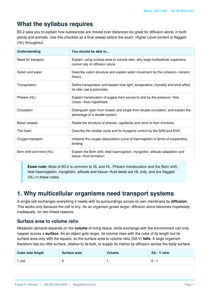
Need for transport Explain, using surface area to volume ratio, why large multicellular organisms cannot rely on diffusion alone.
Xylem and water Describe xylem structure and explain water movement by the cohesion−tension theory.
Transpiration Define transpiration and explain how light, temperature, humidity and wind affect its rate; use a potometer.
Phloem (HL) Explain translocation of sugars from source to sink by the pressure−flow (mass−flow) hypothesis.
Circulation Distinguish open from closed, and single from double circulation, and explain the advantage of a double system.
Blood vessels Relate the structure of arteries, capillaries and veins to their functions.
The heart Describe the cardiac cycle and its myogenic control by the SAN and AVN.
Oxygen transport Interpret the oxygen dissociation curve of haemoglobin in terms of cooperative binding.
Bohr shift and more (HL) Explain the Bohr shift, fetal haemoglobin, myoglobin, altitude adaptation and tissue−fluid formation.
Exam note: Most of B3.2 is common to SL and HL. Phloem translocation and the Bohr shift, fetal haemoglobin, myoglobin, altitude and tissue−fluid detail are HL only, and are flagged (HL) in these notes.
1. Why multicellular organisms need transport systems
A single cell exchanges everything it needs with its surroundings across its own membrane by diffusion. This works only because the cell is tiny. As an organism grows larger, diffusion alone becomes hopelessly inadequate, for two linked reasons.
Surface area to volume ratio
Metabolic demand depends on the volume of living tissue, while exchange with the environment can only happen across a surface. As an object gets larger, its volume rises with the cube of its length but its surface area only with the square, so the surface area to volume ratio (SA:V) falls. A large organism therefore has too little surface, relative to its bulk, to supply its interior by diffusion across the body surface.
Cube side length Surface area Volume SA : V ratio
1 unit 6 : 1
Cube side length Surface area Volume SA : V ratio
2 units 3 : 1
4 units 1.5 : 1
Diffusion is too slow over distance
Diffusion is fast over micrometres but catastrophically slow over millimetres or more, because the time taken is proportional to the square of the distance. A molecule that crosses a cell in a fraction of a second would take hours to diffuse across a whole organ. Deep cells would be starved of oxygen and glucose and poisoned by their own waste long before diffusion could relieve them.
Mass flow: the solution
Large organisms solve the problem with mass flow (bulk flow): the movement of a whole fluid, and everything dissolved or suspended in it, in one direction, driven by a pressure difference. Blood in animals and water in xylem both move by mass flow. This transports substances quickly over long distances; short-range delivery from the transport fluid to individual cells is then completed by diffusion across a large, specialised exchange surface (for example capillary walls or leaf mesophyll).
Key idea: Transport systems do not replace diffusion, they support it. Mass flow covers the long distance quickly; diffusion finishes the last few micrometres across a large surface area with a short diffusion distance and a steep concentration gradient.
2. Transport of water in plants
Plants move water and dissolved minerals upward from the roots to the leaves through the xylem. Remarkably, this happens with no pump: the energy comes from the sun, through evaporation at the leaves.
Structure of xylem
- Xylem vessels are made of dead, empty cells joined end to end. The end walls break down to form a continuous hollow tube, like a drinking straw.
- Because the cells are dead and hollow, there is no cytoplasm or membranes to obstruct flow, giving very low resistance to the moving water column.
- The walls are thickened and waterproofed with lignin, laid down in rings or spirals. Lignin gives mechanical strength, resists the inward pull of tension, and stops the vessel collapsing.
- Xylem also provides structural support for the whole plant (wood is largely old xylem).
Transpiration
Transpiration is the loss of water vapour from the leaves and other aerial parts of a plant, mainly through the stomata of the leaf. Water evaporates from the moist surfaces of the mesophyll cells into the air spaces, then diffuses out through the open stomata down a concentration gradient into the drier air outside. The stomata must open to let CO2 in for photosynthesis, so transpiration is an unavoidable consequence of gas exchange.
The cohesion−tension theory
This theory explains how water is pulled up a tall tree against gravity, in an unbroken column, without a pump.
- Evaporation of water from mesophyll cell surfaces in the leaf lowers the water potential there and creates a tension (negative pressure) at the top of the xylem.
- Cohesion: water molecules are attracted to one another by hydrogen bonding, so they stick together as a continuous column. The tension at the top is transmitted all the way down, pulling the whole column upward.
- Adhesion: water molecules are also attracted to the lignified xylem walls, which helps hold the column in place and resist its weight.
- The result is a continuous transpiration stream pulled from root to leaf. Because the column is under tension, the xylem must be strong (lignin) to avoid collapsing inward.
Root pressure
A second, weaker mechanism operates at the base. Cells in the root actively pump mineral ions into the xylem, lowering its water potential, so water follows by osmosis from the surrounding root cells. This builds up a positive root pressure that pushes water a short way up the stem. Root pressure alone cannot account for water reaching the top of a tall tree; cohesion−tension does the main work, especially when transpiration is rapid.
Factors affecting the rate of transpiration
Anything that steepens the water−vapour gradient between the leaf air spaces and the outside air, or speeds evaporation, increases the transpiration rate.
Factor Effect on rate Explanation
Light intensity Increase Light causes stomata to open for photosynthesis; more open stomata means more evaporation.
Temperature Increase Warmer air gives water molecules more kinetic energy, so evaporation is faster and the air can hold more vapour, steepening the gradient.
Humidity Decrease Humid air already holds much water vapour, so the concentration gradient out of the leaf is shallower and evaporation is slower.
Wind / air movement Increase Moving air carries away vapour that would otherwise accumulate near the stomata, keeping the gradient steep.
Measuring transpiration: the potometer
A potometer measures the rate at which a cut shoot takes up water, which closely tracks the transpiration rate. A leafy shoot is sealed into a water−filled tube connected to a capillary tube with an air bubble. As the shoot transpires, water is drawn in and the bubble moves along a scale; the distance moved per unit time gives the rate. A reservoir tap lets you reset the bubble.
Strictly, a potometer measures water uptake, not water loss; the two differ slightly because a little water is used in photosynthesis and to keep cells turgid. To make results comparable, change only one factor at a time (for example use a fan for wind, or a lamp for light) and keep the others constant.
Common misconception: Water is not pushed up from below by the roots in most plants. It is pulled up from above by tension generated by evaporation at the leaves. The column is under negative pressure, which is why a cut stem draws air in rather than squirting water out.
3. Transport of sugars in plants (HL)
Water rises in the xylem, but the products of photosynthesis must be distributed in all directions, to wherever they are needed. This is the job of the phloem, and the process is called translocation. (HL)
Phloem structure (HL)
- Sieve tube elements are living cells joined end to end. Their end walls are perforated to form sieve plates, letting sap flow through.
- To reduce resistance to flow, sieve tube elements lose their nucleus and most organelles at maturity, so they depend on neighbouring companion cells.
- Companion cells are dense with mitochondria and provide the ATP for the active loading of sugar into the phloem.
Source and sink (HL)
A source is any part that loads sugar into the phloem, typically a photosynthesising leaf or a storage organ that is exporting. A sink is any part that removes sugar, such as a growing root tip, a developing fruit or a storage organ that is filling. Translocation runs from source to sink, so its direction can change with the season: a potato tuber is a sink while it fills in summer, then a source when it sprouts in spring.
The pressure−flow (mass−flow) hypothesis (HL)
Translocation is explained by the pressure−flow hypothesis, which links active loading at the source to bulk flow driven by a pressure gradient.
- At the source, sucrose is actively loaded into the sieve tubes using ATP from the companion cells. This lowers the water potential of the phloem sap.
- Water follows by osmosis from the nearby xylem into the phloem, raising the hydrostatic pressure at the source end.
- At the sink, sucrose is unloaded (used or stored), water leaves the phloem by osmosis, and the hydrostatic pressure falls.
- The pressure difference between source (high) and sink (low) drives phloem sap by mass flow from source to sink, carrying the dissolved sugars with it.
Contrast to remember (HL): Xylem transport is passive, one−directional (up), through dead cells, driven by evaporation. Phloem transport needs ATP for loading, is bidirectional (source to sink), through living cells, driven by a hydrostatic pressure gradient.
Figure 1. Xylem transport (transpiration stream: one-way, upward, in dead lignified vessels) alongside phloem translocation
(HL: source → sink mass flow in living sieve tubes).
4. Animal circulatory systems
Animals large enough to need a transport system move a fluid (blood or haemolymph) around the body by a muscular pump. Circulatory systems are classified in two ways.
Open versus closed
Open circulation Closed circulation
Where fluid goes Pumped into open body cavities (haemocoel); bathes organs directly
Confined to a continuous network of vessels
Pressure Low High, so transport is faster
Exchange Directly with tissues Across capillary walls; flow to organs can be regulated
Found in Most arthropods and molluscs Annelids and all vertebrates
Single versus double circulation
In a single circulation (for example a fish), blood passes through the heart once per complete circuit: heart → gills → body → heart. Blood loses much of its pressure passing through the tiny gill capillaries, so it flows to the body relatively slowly.
In a double circulation (for example a mammal), blood passes through the heart twice per circuit. The heart is divided into two pumps side by side:
- Pulmonary circuit: right side of the heart → lungs → back to the left side of the heart.
- Systemic circuit: left side of the heart → rest of the body → back to the right side of the heart.
Why a double circulation is efficient
Blood pressure drops steeply on passing through any capillary bed. In a double system the blood returns to the heart after the lungs and is re-pressurised before being sent to the body, so it reaches the tissues at high pressure and flows quickly. This supports the high metabolic rate of a warm−blooded mammal. It also keeps oxygenated and deoxygenated blood fully separated, so the tissues receive blood with the maximum oxygen content.
Exam tip: The right side of the heart handles deoxygenated blood going to the lungs; the left side handles oxygenated blood going to the body. The left ventricle wall is thicker because it must generate the higher pressure needed to reach the whole body.
Figure 2. Mammalian double circulation: right heart → pulmonary circuit → left heart → systemic circuit → right heart. Blood passes through the heart twice per full circuit.
5. Blood vessels
Blood leaves the heart in arteries, which branch into narrower arterioles, then into capillaries where exchange occurs. Capillaries join into venules, which merge into veins that return blood to the heart. Each vessel type is built for its role.
Feature Arteries Capillaries Veins
Direction Carry blood away from the heart Link arterioles to venules Carry blood back to the heart
Wall Thick, with muscle and elastic tissue
One cell thick (endothelium only)
Thin, less muscle and elastic tissue
Lumen Narrow Very narrow (one red cell wide) Wide
Pressure High and pulsing Falling Low
Valves None (except at heart exit) None Present, to prevent backflow
How the structures fit the functions
- Arteries: the thick, elastic, muscular wall withstands and smooths the high, pulsing pressure. The wall stretches as the heart pumps, then recoils, keeping blood moving between beats and evening out the flow.
- Capillaries: a wall just one cell thick gives a very short diffusion distance; the huge total number provides an enormous surface area, and the narrow lumen slows the blood, giving time for exchange of O2, CO2, glucose and wastes.
- Veins: a wide lumen offers little resistance to low−pressure return flow. Because pressure is too low to keep blood moving upward, valves prevent backflow and contraction of nearby skeletal muscles squeezes the veins to push blood back toward the heart.
Common misconception: Arteries do not always carry oxygenated blood. They carry blood away from the heart. The pulmonary artery carries deoxygenated blood to the lungs, and the pulmonary vein carries oxygenated blood back. Define arteries and veins by direction, not by oxygen content.
6. The heart and the cardiac cycle
The mammalian heart is a double pump with four chambers: two thin−walled atria on top that receive blood, and two thick−walled ventricles below that pump it out. The muscular septum separates the two sides so oxygenated and deoxygenated blood never mix.
Valves keep blood flowing one way
- Atrioventricular (AV) valves between each atrium and ventricle prevent backflow into the atria when the ventricles contract.
- Semilunar valves at the exits into the aorta and pulmonary artery prevent backflow into the ventricles when they relax.
The cardiac cycle
One heartbeat is one cardiac cycle, a repeating sequence of contraction (systole) and relaxation (diastole).
Stage What happens Valves
Atrial systole Atria contract, pushing the last of the blood into the relaxed ventricles. AV valves open; semilunar closed
Ventricular systole Ventricles contract; pressure rises and blood is forced out into the aorta and pulmonary artery.
AV valves close; semilunar open
Diastole Atria and ventricles relax; blood flows into the atria and passively fills the ventricles.
Semilunar closed; AV valves open
Myogenic contraction and its control
Cardiac muscle is myogenic: it contracts and relaxes rhythmically on its own, without a signal from the nervous system. The rhythm is set and coordinated by electrical activity within the heart itself.
- The sinoatrial node (SAN) in the wall of the right atrium is the pacemaker. It fires a wave of electrical excitation that spreads across both atria, making them contract.
- The excitation reaches the atrioventricular node (AVN), which imposes a brief delay, so the atria finish emptying before the ventricles contract.
- The AVN then passes the signal down conducting fibres to the apex of the ventricles, so they contract from the bottom up, pushing blood upward into the arteries.
The intrinsic rate can be adjusted by the nervous system and by hormones such as adrenaline, which speed the heart during exercise or stress.
Heart sounds and blood pressure
The familiar “lub−dub” heard through a stethoscope is the sound of valves closing: the first sound is the AV valves closing at the start of ventricular systole, the second is the semilunar valves closing at the start of diastole. Blood pressure is usually quoted as two numbers, for example 120/80: the higher systolic pressure during ventricular contraction over the lower diastolic pressure while the heart relaxes.
Exam tip: Learn the order: atrial systole → ventricular systole → diastole. Link each valve movement to the pressure change that causes it. A valve opens when the pressure behind it exceeds the pressure in front, and closes when the pressure in front becomes greater.
Figure 3. Cardiac cycle pressure-time graph (HR = 75 bpm, T = 0.8 s). The semilunar (SL) valve opens and closes where ventricular pressure crosses the aortic diastolic pressure (80 mmHg), computed from sin(πx) = (Pdia−P0)/(Psys−P0).
7. Transport of oxygen: haemoglobin
Oxygen is only slightly soluble in plasma, so almost all of it is carried by haemoglobin inside red blood cells. Each haemoglobin molecule has four subunits, each with an iron−containing haem group that binds one O2 molecule, so one haemoglobin can carry up to four O2.
Loading and unloading
Haemoglobin must do two opposite jobs: pick up oxygen where it is plentiful and release it where it is scarce. It manages both because its affinity for oxygen changes with the surrounding oxygen concentration, measured as the partial pressure of oxygen (pO2).
- In the lungs pO2 is high, haemoglobin has a high affinity for oxygen and loads (becomes almost fully saturated).
- In respiring tissues pO2 is low, affinity is lower and haemoglobin unloads oxygen for the cells to use.
The oxygen dissociation curve
A graph of percentage saturation of haemoglobin (y−axis) against pO2 (x−axis) is the oxygen dissociation curve. It is S−shaped (sigmoid), and this shape is the key to how haemoglobin works.
- The shape arises from cooperative binding: when the first O2 binds, it slightly changes the shape of the haemoglobin molecule, making it easier for the next O2 to bind. Affinity rises as each O2 loads.
- This makes the curve steep in the middle: over the range of pO2 found in the tissues, a small fall in pO2 causes a large release of oxygen, delivering plenty of O2 exactly where respiration is fastest.
- The curve flattens at high pO2, so haemoglobin still loads almost fully in the lungs even if lung pO2 dips a little.
Reading the curve: A curve shifted to the left means a higher affinity (loads oxygen more readily, unloads less). A curve shifted to the right means a lower affinity (unloads oxygen more readily). Keep this “left = loads, right = releases” rule ready for the Bohr shift.
8. Fine control of oxygen delivery (HL)
Higher Level extends the dissociation curve to show how oxygen delivery is tuned to need, and how oxygen leaves the blood to reach cells. (HL)
The Bohr shift (HL)
Actively respiring tissue produces CO2, which lowers the pH of the blood (it forms carbonic acid). A rise in CO2 and fall in pH lowers haemoglobin's affinity for oxygen, shifting the dissociation curve to the right. This is the Bohr shift. Its benefit is elegant: the tissues that are respiring hardest, and so producing the most CO2, are exactly the tissues that most need oxygen, and the Bohr shift makes haemoglobin release more oxygen there, at any given pO2. (HL)
Fetal haemoglobin (HL)
A fetus must obtain oxygen from its mother's blood across the placenta. Fetal haemoglobin has a higher affinity for oxygen than adult haemoglobin, so its dissociation curve lies to the left of the adult curve. At the placenta, therefore, fetal haemoglobin can take up oxygen that maternal haemoglobin is releasing, allowing net transfer of oxygen from mother to fetus. (HL)
Myoglobin (HL)
Myoglobin is an oxygen−binding pigment in muscle. It has a very high affinity for oxygen (its curve is far to the left and is not sigmoid but hyperbolic, because it has a single subunit and no cooperativity). Myoglobin only releases its oxygen at the very low pO2 of hard exercise, so it acts as an oxygen store that is called on when demand is extreme. (HL)
Adaptation to high altitude (HL)
At high altitude the pO2 of the air is low, so it is harder to load oxygen in the lungs. People living at altitude (and those who acclimatise) adapt in several ways: they produce more red blood cells and more haemoglobin, raising the oxygen−carrying capacity of the blood; they may have a higher breathing rate; and they develop a greater density of capillaries so oxygen reaches the tissues more effectively. Some high−altitude species also have haemoglobin with a higher oxygen affinity. (HL)
Capillary exchange and tissue fluid (HL)
Exchange between blood and cells happens through tissue fluid, the fluid that bathes the cells. It forms and drains as blood passes along a capillary. (HL)
- At the arterial end, the high hydrostatic (blood) pressure exceeds the osmotic pull of the plasma proteins, so fluid is forced out of the capillary by ultrafiltration. Water, oxygen, glucose and ions leave; large plasma proteins and cells are held back.
- This fluid, now tissue fluid, bathes the cells and lets them exchange substances with the blood by diffusion.
- Along the capillary, hydrostatic pressure falls while the plasma proteins left behind keep the blood's osmotic pull high. At the venous end the osmotic pull now exceeds the hydrostatic pressure, so much of the fluid, plus CO2 and other wastes, is drawn back into the capillary.
- The surplus fluid that does not return is collected by the lymphatic system and eventually returned to the blood.
Exam tip (HL): Tissue fluid formation is a balance of two pressures: outward hydrostatic (blood) pressure versus inward osmotic pressure from plasma proteins. Net outflow at the arterial end, net inflow at the venous end. The lymphatic system mops up the difference.
9. Worked examples and exam skills
Worked example 1 - explaining cohesion−tension
Explain how water moves from the roots to the leaves of a tall tree. [4 marks]
Water evaporates from the mesophyll cells and diffuses out of the stomata (transpiration), creating a tension at the top of the xylem (1). Water molecules are held together in a continuous column by cohesion (hydrogen bonding) (1), so the tension pulls the whole column upward (1). Adhesion to the xylem walls helps support the column against gravity (1).
Worked example 2 - interpreting potometer data
A potometer bubble moved 60 mm in 5 minutes in still air, then 150 mm in 5 minutes when a fan was switched on. The capillary tube cross−section is 1.0 mm2. Find each rate of water uptake and explain the difference.
Still air: 60 × 1.0 = 60 mm3 in 5 min = 12 mm3 min−1. With fan: 150 × 1.0 = 150 mm3 in 5 min = 30 mm3 min−1.
The fan increases air movement, which carries away water vapour from around the stomata, keeping the concentration gradient out of the leaf steep, so evaporation and hence uptake are faster.
Worked example 3 - reading an oxygen dissociation curve
At the lungs pO2 is about 13 kPa and haemoglobin is 98% saturated; in an exercising muscle pO2 is about 3 kPa and saturation is 30%. What percentage of the oxygen carried is unloaded to the muscle, and why is so much released?
Oxygen unloaded = 98 − 30 = 68% of capacity is released to the muscle.
Because the curve is steep in this middle region, a small drop in pO2 causes a large fall in saturation, so a large amount of oxygen is released exactly where respiration is fastest. Added CO2 shifts the curve right (Bohr shift), releasing even more (HL).
Worked example 4 - why a double circulation is efficient
Explain one advantage of a double circulation over a single circulation. [3 marks]
In a single circulation, blood loses pressure passing through the gill capillaries before reaching the body, so it flows slowly (1). In a double circulation the blood returns to the heart and is re-pressurised after the lungs (1), so it travels to the tissues at high pressure and delivers oxygen quickly, supporting a high metabolic rate (1).
Worked example 5 - ordering the cardiac cycle
Put these events in order and state the valve positions: ventricles contract; atria contract; heart relaxes.
- Atrial systole - atria contract, AV valves open, semilunar closed. 2. Ventricular systole - ventricles contract, AV valves close (first heart sound), semilunar open. 3. Diastole - all relax, semilunar close (second heart sound), AV valves open and ventricles refill.
Common pitfalls to avoid
- Xylem is dead; phloem is living. Xylem vessels are dead, hollow, lignified tubes; phloem sieve tubes are living cells with companion cells. Do not mix them up.
- Transpiration is not translocation. Transpiration is water loss and water movement in the xylem; translocation is sugar movement in the phloem (HL).
- Arteries carry blood away from the heart, veins carry it back, regardless of oxygen content (remember the pulmonary vessels).
- Water is pulled up the xylem by tension, not pushed up from the roots; root pressure plays only a minor part.
- A left−shifted dissociation curve means higher affinity (loads oxygen); a right−shifted curve means lower affinity (releases oxygen).
- Define systole as contraction and diastole as relaxation, and give valve positions in terms of the pressure differences that cause them.
Quick reference
Idea Statement
Need for transport SA:V falls as size rises; diffusion time ≈ (distance)2; mass flow solves the long−distance problem.
Cohesion−tension Evaporation at leaves → tension; cohesion holds the column; adhesion supports it; water is pulled up.
Transpiration factors Light, temperature and wind raise the rate; humidity lowers it.
Phloem (HL) Active loading at source → water in by osmosis → high pressure → mass flow to sink.
Double circulation Heart re-pressurises blood after the lungs; oxygenated and deoxygenated blood kept separate.
Vessels Arteries: thick, elastic, high pressure. Capillaries: one cell thick, exchange. Veins: wide lumen, valves.
Cardiac cycle Atrial systole → ventricular systole → diastole; SAN paces, AVN delays.
Dissociation curve Sigmoid; cooperative binding; steep in tissues (unloads), flat in lungs (loads).
Bohr shift (HL) More CO2 / lower pH → curve shifts right → more O2 released.
10. Test yourself
Attempt all eight questions without notes, then check against the full answers below.
- Explain, in terms of surface area to volume ratio, why an elephant needs a transport system but a single−celled organism does not.
- Describe two features of xylem vessels and relate each to its function.
- A student wants to investigate the effect of wind on transpiration using a potometer. State the independent variable and two variables that must be controlled.
- (HL) Outline how sucrose is moved from a leaf to a growing root by the pressure−flow hypothesis.
- Explain why the wall of the left ventricle is thicker than that of the right ventricle.
- Describe the roles of the SAN and the AVN in controlling the heartbeat.
- Explain how the sigmoid shape of the oxygen dissociation curve helps haemoglobin deliver oxygen to respiring tissue.
- (HL) Explain the Bohr shift and why it is useful during exercise.
Answers
- A single−celled organism has a large SA:V, so diffusion across its surface supplies its whole small volume fast enough. An elephant has a small SA:V and a large diffusion distance to inner cells, so diffusion alone is far too slow; mass flow in a transport system is needed.
- Vessels are dead and hollow (no cytoplasm), giving a continuous low−resistance tube for water flow; walls are strengthened with lignin, which provides support and stops the vessel collapsing under the tension of the water column.
- Independent variable: air movement / wind speed (for example fan setting). Control at least two of: temperature, light intensity, humidity, the same shoot / leaf area, and time interval.
- (HL) At the leaf (source) sucrose is actively loaded into the phloem using ATP from companion cells, lowering the water potential; water enters by osmosis, raising hydrostatic pressure. At the root (sink) sucrose is unloaded and water leaves, lowering pressure. The pressure gradient drives phloem sap by mass flow from source to sink.
- The left ventricle pumps blood around the whole body (systemic circuit) and must generate a much higher pressure than the right ventricle, which only pumps to the nearby lungs; a thicker, more muscular wall produces this greater force.
- The SAN (pacemaker) in the right atrium initiates each heartbeat, sending excitation across the atria to make them contract. The AVN receives this, delays it briefly so the atria empty first, then passes it to the ventricle apex so the ventricles contract from the bottom up.
- Cooperative binding makes the curve steep in the middle, over the pO2 range found in tissues, so a small fall in pO2 there causes a large drop in saturation and a large release of oxygen where it is needed. The flat top means haemoglobin still loads fully in the lungs.
- (HL) Rising CO2 lowers blood pH, which reduces haemoglobin's affinity for oxygen and shifts the curve to the right, so at any given pO2 more oxygen is unloaded. During exercise, hard−working muscle produces more CO2 and so receives more oxygen, exactly where demand is highest.
