
A Level · Biology 9700 · Gas exchange: summary notes
Gas exchange: summary notes
Opens inside this page. Prefer your own PDF app? Open the PDF.
Check yourself
5 quick questions on A Level Biology
Tap an answer to see if you are right, and why. Free, no sign-up.
Start the free test12 of 12 students found these notes useful
Was this note useful?
Full text of Gas exchange: summary notes
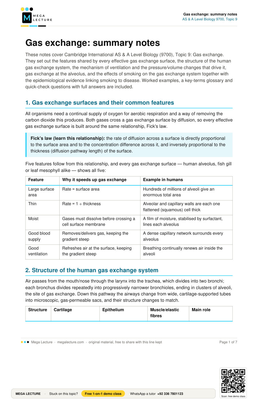
These notes cover Cambridge International AS & A Level Biology (9700), Topic 9: Gas exchange. They set out the features shared by every effective gas exchange surface, the structure of the human gas exchange system, the mechanism of ventilation and the pressure/volume changes that drive it, gas exchange at the alveolus, and the effects of smoking on the gas exchange system together with the epidemiological evidence linking smoking to disease. Worked examples, a key-terms glossary and quick-check questions with full answers are included.
1. Gas exchange surfaces and their common features
All organisms need a continual supply of oxygen for aerobic respiration and a way of removing the carbon dioxide this produces. Both gases cross a gas exchange surface by diffusion, so every effective gas exchange surface is built around the same relationship, Fick's law.
Fick's law (learn this relationship): the rate of diffusion across a surface is directly proportional to the surface area and to the concentration difference across it, and inversely proportional to the thickness (diffusion pathway length) of the surface.
Five features follow from this relationship, and every gas exchange surface — human alveolus, fish gill or leaf mesophyll alike — shows all five:
| Feature | Why it speeds up gas exchange | Example in humans |
|---|---|---|
| Large surface area | Rate ∝ surface area | Hundreds of millions of alveoli give an enormous total area |
| Thin | Rate ∝ 1 ÷ thickness | Alveolar and capillary walls are each one flattened (squamous) cell thick |
| Moist | Gases must dissolve before crossing a cell surface membrane | A film of moisture, stabilised by surfactant, lines each alveolus |
| Good blood supply | Removes/delivers gas, keeping the gradient steep | A dense capillary network surrounds every alveolus |
| Good ventilation | Refreshes air at the surface, keeping the gradient steep | Breathing continually renews air inside the alveoli |
2. Structure of the human gas exchange system
Air passes from the mouth/nose through the larynx into the trachea, which divides into two bronchi; each bronchus divides repeatedly into progressively narrower bronchioles, ending in clusters of alveoli, the site of gas exchange. Down this pathway the airways change from wide, cartilage-supported tubes into microscopic, gas-permeable sacs, and their structure changes to match.
| Structure | Cartilage | Epithelium | Muscle/elastic fibres | Main role |
|---|---|---|---|---|
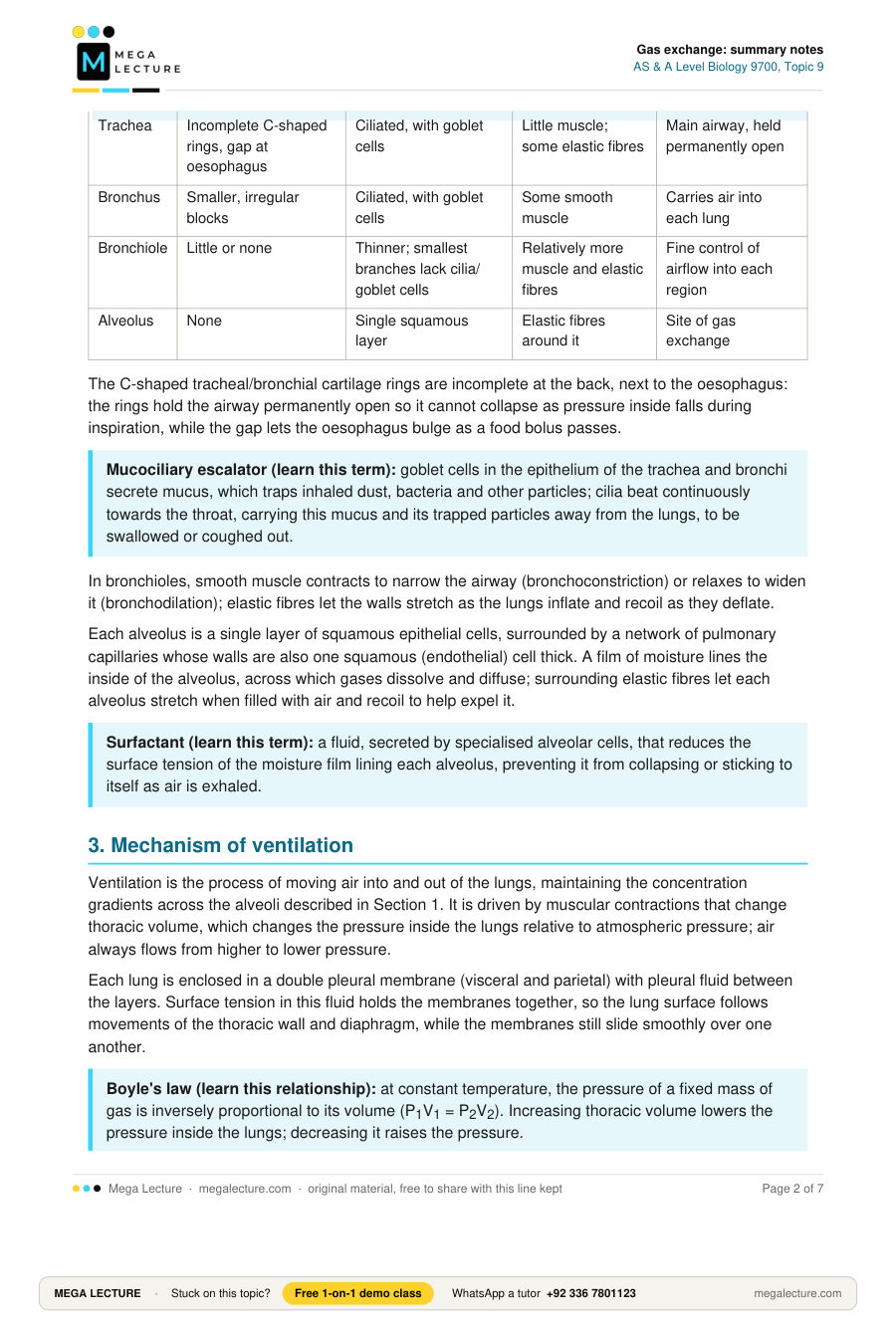
| Trachea | Incomplete C-shaped rings, gap at oesophagus | Ciliated, with goblet cells | Little muscle; some elastic fibres | Main airway, held permanently open |
| Bronchus | Smaller, irregular blocks | Ciliated, with goblet cells | Some smooth muscle | Carries air into each lung |
| Bronchiole | Little or none | Thinner; smallest branches lack cilia/goblet cells | Relatively more muscle and elastic fibres | Fine control of airflow into each region |
| Alveolus | None | Single squamous layer | Elastic fibres around it | Site of gas exchange |
The C-shaped tracheal/bronchial cartilage rings are incomplete at the back, next to the oesophagus: the rings hold the airway permanently open so it cannot collapse as pressure inside falls during inspiration, while the gap lets the oesophagus bulge as a food bolus passes.
Mucociliary escalator (learn this term): goblet cells in the epithelium of the trachea and bronchi secrete mucus, which traps inhaled dust, bacteria and other particles; cilia beat continuously towards the throat, carrying this mucus and its trapped particles away from the lungs, to be swallowed or coughed out.
In bronchioles, smooth muscle contracts to narrow the airway (bronchoconstriction) or relaxes to widen it (bronchodilation); elastic fibres let the walls stretch as the lungs inflate and recoil as they deflate.
Each alveolus is a single layer of squamous epithelial cells, surrounded by a network of pulmonary capillaries whose walls are also one squamous (endothelial) cell thick. A film of moisture lines the inside of the alveolus, across which gases dissolve and diffuse; surrounding elastic fibres let each alveolus stretch when filled with air and recoil to help expel it.
Surfactant (learn this term): a fluid, secreted by specialised alveolar cells, that reduces the surface tension of the moisture film lining each alveolus, preventing it from collapsing or sticking to itself as air is exhaled.
3. Mechanism of ventilation
Ventilation is the process of moving air into and out of the lungs, maintaining the concentration gradients across the alveoli described in Section 1. It is driven by muscular contractions that change thoracic volume, which changes the pressure inside the lungs relative to atmospheric pressure; air always flows from higher to lower pressure.
Each lung is enclosed in a double pleural membrane (visceral and parietal) with pleural fluid between the layers. Surface tension in this fluid holds the membranes together, so the lung surface follows movements of the thoracic wall and diaphragm, while the membranes still slide smoothly over one another.
Boyle's law (learn this relationship): at constant temperature, the pressure of a fixed mass of gas is inversely proportional to its volume (P1V1 = P2V2). Increasing thoracic volume lowers the pressure inside the lungs; decreasing it raises the pressure.
Inspiration (always active):
- External intercostal muscles contract, internal intercostals relax, pulling the rib cage up and out.
- The diaphragm contracts, flattening and moving down from its resting dome shape.
- Thoracic volume increases; lung volume increases with it via the pleural membranes and fluid.
- By Boyle's law, alveolar pressure falls below atmospheric pressure.
- Air flows in down the pressure gradient until the two pressures are equal.
Expiration at rest (normally passive):
- External intercostals relax; the rib cage falls under gravity and elastic recoil of stretched lung tissue.
- The diaphragm relaxes, pushed back into its dome shape by the abdominal organs.
- Thoracic and lung volume decrease.
- By Boyle's law, alveolar pressure rises above atmospheric pressure.
- Air flows out down the pressure gradient until pressures equalise.
Forced expiration (active, e.g. exercise or speech): internal intercostals contract, pulling the rib cage down and in more forcefully, while external intercostals relax; abdominal muscles may also contract, pushing the diaphragm further up. This produces a larger, faster fall in thoracic volume than passive recoil alone.
Worked example: At the start of inspiration a person's lung volume is 2.50 dm3 at atmospheric pressure, 101 kPa. Muscle contraction increases lung volume to 2.60 dm3. Use Boyle's law (P1V1 = P2V2) to find the new pressure, and state the direction air flows.
P2 = (P1 × V1) ÷ V2 = (101 × 2.50) ÷ 2.60 = 97.1 kPa (3 s.f.).
97.1 kPa is below atmospheric pressure, so air flows in, from the higher pressure outside to the lower pressure inside, until the pressures equalise.
Worked example: A resting adult has a tidal volume of 500 cm3 and breathes 14 times per minute. Calculate the pulmonary ventilation rate in dm3 per minute.
Pulmonary ventilation rate = tidal volume × breathing rate = 500 × 14 = 7000 cm3 min-1 = 7 dm3 min-1.
4. Gas exchange at the alveolus
Oxygen diffuses from the alveolar air space, through the moisture film lining the alveolus, across the single-celled alveolar epithelium and fused basement membranes, across the single-celled capillary endothelium, into the plasma and then into a red blood cell, where most of it binds to haemoglobin. Carbon dioxide diffuses the opposite way along the same short pathway, into the alveolar air space, removed each time air is exhaled.
This is efficient for the reasons in Section 1 applied here: every layer crossed is only one cell thick (or less, for the basement membranes), giving a very short diffusion pathway; the huge number of alveoli gives an enormous total surface area; and blood flow and ventilation continually renew the gas on each side, keeping both gradients steep across the whole surface.
5. Effects of smoking on the gas exchange system
Tobacco smoke contains thousands of chemicals. Four components matter most here: tar, carbon monoxide, nicotine, and other irritant particulates that damage the ciliated epithelium. Together their long-term effects cause the three principal smoking-related diseases: chronic bronchitis, emphysema and lung cancer.
| Component | What it does | Main long-term effect |
|---|---|---|
| Tar | Carcinogenic mixture (e.g. polycyclic aromatic hydrocarbons); also a general irritant | Mutations in epithelial cells; stimulates excess mucus |
| Carbon monoxide | Binds haemoglobin, forming carboxyhaemoglobin, with far higher affinity than oxygen | Lowers blood's oxygen-carrying capacity; heart works harder |
| Nicotine | Addictive stimulant, absorbed rapidly via the alveoli | Raises heart rate/blood pressure via adrenaline; sustains addiction |
| Other particulates/gases | Paralyse, then destroy, cilia | Removes the mucociliary escalator |
Chronic bronchitis (learn this term): long-term inflammation and narrowing of the bronchi and bronchioles, caused by an irritant such as tobacco smoke, in which goblet cells increase in number and mucus output while ciliary damage stops this mucus being cleared, causing a persistent, mucus-producing cough and repeated infection.
With the mucociliary escalator disabled, the excess mucus accumulates instead of being cleared, providing conditions for bacterial infection; coughing becomes the only remaining way to shift it.
Emphysema (learn this term): destruction of the walls between alveoli by the enzyme elastase, released from phagocytes drawn in by chronic inflammation; this reduces the gas exchange surface area and destroys elastic recoil, causing breathlessness.
Elastase breaks down elastin, the elastic protein in alveolar walls, and smoke also weakens the natural inhibitor that normally limits elastase activity. Alveolar walls merge into fewer, larger spaces with much less surface area, and the remaining tissue can no longer recoil to expel air efficiently, so air is trapped and less oxygen reaches the blood for a given effort — especially noticeable during exercise.
Lung cancer: carcinogens in tar mutate genes that control the cell cycle in bronchial/alveolar epithelial cells, causing uncontrolled mitosis and a tumour that may invade surrounding tissue and metastasise via the blood or lymphatic system.
6. Epidemiological evidence linking smoking to disease
Epidemiology (learn this term): the study of the patterns, causes and effects of disease in populations, used to identify factors, such as smoking, associated with increased disease risk.
Since it would be unethical to expose people to smoke experimentally, the case for smoking causing disease rests on large-scale observational studies. Several strands of evidence together build a case for causation:
- Correlation: long-term cohort studies consistently find a higher incidence of lung cancer, chronic bronchitis, emphysema and cardiovascular disease among smokers.
- Dose-response relationship: risk rises with cigarettes smoked per day and years smoked ("pack-years").
- Consistency: the same association recurs across many independent studies and populations.
- Reversibility: excess risk falls progressively after smokers quit, supporting a causal (not just correlational) link.
- Biological plausibility: known mechanisms (mutagens in tar, elastase in emphysema, ciliary damage in bronchitis) explain the pattern seen.
Because the evidence is observational, researchers must control for confounding variables — factors such as occupational pollutant exposure or diet that could independently affect disease risk and also be commoner among smokers. Well-designed cohort studies adjust for these statistically, strengthening the causal conclusion.
Worked example: In a cohort study, 1200 per 100 000 smokers developed lung cancer, versus 60 per 100 000 non-smokers. Calculate the relative risk for a smoker (illustrative data).
Relative risk = incidence in exposed ÷ incidence in unexposed = (1200 ÷ 100 000) ÷ (60 ÷ 100 000) = 20.
A smoker here was 20 times as likely as a non-smoker to develop lung cancer — strong supporting evidence, though relative risk alone does not prove causation (see above).
Key terms
| Term | Definition |
|---|---|
| Gas exchange surface | A surface across which respiratory gases diffuse between organism and environment |
| Fick's law | Diffusion rate ∝ surface area × concentration difference ÷ diffusion distance |
| Ventilation | Moving air into and out of the lungs by muscular contraction |
| Trachea | Main airway, held open by incomplete cartilage rings |
| Bronchus | Airway connecting the trachea to a lung, supported by cartilage |
| Bronchiole | Fine airway with little/no cartilage but relatively more smooth muscle |
| Alveolus | Tiny air sac at the end of a bronchiole; site of gas exchange |
| Ciliated epithelium | Epithelium bearing cilia that move mucus along the airway |
| Goblet cell | Cell in the airway epithelium that secretes mucus |
| Mucociliary escalator | Beating cilia moving mucus and trapped particles out of the airways |
| Squamous epithelium | A single layer of thin, flattened cells, as in the alveolar wall |
| Surfactant | Fluid that lowers surface tension in the alveolus, preventing collapse |
| Pleural membrane | One of two membranes enclosing each lung, with fluid between them |
| Intercostal muscles | Muscles between ribs; external/internal sets act antagonistically |
| Diaphragm | Dome-shaped muscle sheet separating thorax from abdomen |
| Boyle's law | For a fixed mass of gas at constant temperature, pressure ∝ 1 ÷ volume |
| Tidal volume | Volume of air moved in one normal breath |
| Pulmonary ventilation rate | Tidal volume × breathing rate; air breathed per minute |
| Tar | Carcinogenic mixture in smoke that also irritates the airways |
| Carboxyhaemoglobin | Compound formed when carbon monoxide binds haemoglobin |
| Nicotine | Addictive stimulant in smoke that raises heart rate/blood pressure |
| Chronic bronchitis | Long-term inflamed, narrowed, mucus-filled bronchi/bronchioles |
| Emphysema | Destruction of alveolar walls by elastase, reducing exchange surface |
| Elastase | Enzyme, from phagocytes, that breaks down elastin |
| Epidemiology | Study of disease patterns, causes and effects in populations |
| Confounding variable | A factor other than the one studied that could independently affect the outcome |
| Relative risk | Incidence in an exposed group ÷ incidence in an unexposed group |
Quick check
- State two features shared by all efficient gas exchange surfaces. [2]
- Define the term "ventilation". [1]
- Describe the role of the cartilage in the trachea. [2]
- Describe three ways a bronchiole's structure differs from a bronchus's. [3]
- Explain how the alveolar and capillary walls are adapted for efficient gas exchange. [4]
- Explain, in terms of pressure and volume, how the diaphragm and external intercostal muscles bring about inspiration. [5]
- Explain why expiration at rest is passive, while forced expiration is not. [3]
- Calculate the pulmonary ventilation rate for a tidal volume of 450 cm3 and a breathing rate of 16 min-1. Give your answer in dm3 min-1. [2]
- Explain how smoking can lead to chronic bronchitis. [4]
- Explain how smoking can lead to emphysema, and why a person with emphysema becomes breathless on exertion. [5]
- A study found smokers several times more likely to develop lung cancer than non-smokers. Give two reasons why correlation alone does not prove causation here, and suggest two further types of evidence that would strengthen the case. [4]
- Explain why a smoker's gas exchange system is more vulnerable to respiratory infection than a non-smoker's. [4]
1. Any two of: large surface area; thin; moist; good blood supply; good ventilation.
2. Moving air into and out of the lungs by muscular contraction, changing thoracic (and so alveolar) pressure.
3. C-shaped rings hold the trachea open so it cannot collapse as internal pressure falls in inspiration; the gap, next to the oesophagus, lets it bulge as food passes.
4. Any three of: bronchioles have little/no cartilage vs cartilage in bronchi; proportionally more smooth muscle and elastic fibres, allowing constriction/dilation and stretch/recoil; narrower diameter; smallest bronchioles lack cilia/goblet cells.
5. Both are a single squamous cell thick, giving a short diffusion pathway; the huge alveolar/capillary number gives a large surface area; the surface is moist so gases dissolve; the dense capillary network maintains a steep gradient by removing/supplying gas.
6. External intercostal contraction pulls the rib cage up/out; diaphragm contraction flattens it downward; both increase thoracic volume, and lung volume with it (pleural membranes/fluid); by Boyle's law this fall in pressure takes alveolar pressure below atmospheric; air flows in down the gradient until pressures equalise.
7. At rest, elastic recoil and gravity are enough to reduce thoracic volume; forced expiration must expel air faster/more completely, so internal intercostal and abdominal muscles contract to reduce thoracic volume actively.
8. 450 × 16 = 7200 cm3 min-1 = 7.2 dm3 min-1.
9. Smoke damages/destroys cilia while goblet cells increase mucus output; with the mucociliary escalator disabled, mucus accumulates, narrowing airways, causing inflammation and repeated infection; coughing becomes the only way to clear it.
10. Chronic inflammation draws in phagocytes, which release elastase while digesting debris; elastase breaks down elastin (its natural inhibitor is also weakened by smoke); alveolar walls merge into fewer, larger spaces with less surface area and lost elastic recoil; air becomes trapped and less oxygen reaches the blood per effort, so breathlessness appears, especially on exertion.
11. Any two of: correlation alone cannot rule out a confounding variable independently raising cancer risk and being commoner among smokers; correlation does not establish a mechanism, or the direction of cause and effect, linking the two variables. Any two of: a dose-response relationship; consistency across independent studies/populations; falling risk after quitting; a known biological mechanism linking smoke chemicals to the mutations seen.
12. Tar and other particulates in tobacco smoke paralyse and then destroy the cilia in the epithelium lining the trachea and bronchi, disabling the mucociliary escalator; goblet cells also increase in number and mucus output in response to the irritants. With cilia no longer able to clear it, this excess mucus — along with trapped bacteria and other particles — accumulates in the airways instead of being swallowed or coughed away, so pathogens are retained in the airways for longer, increasing the risk of infection.
