
A Level · Biology 9700 · Biological molecules: summary notes
Biological molecules: summary notes
Opens inside this page. Prefer your own PDF app? Open the PDF.
Check yourself
5 quick questions on A Level Biology
Tap an answer to see if you are right, and why. Free, no sign-up.
Start the free test12 of 12 students found these notes useful
Was this note useful?
Full text of Biological molecules: summary notes
These revision notes cover Topic 2 (Biological molecules) of the Cambridge International AS & A Level Biology syllabus (9700). They follow the topic in syllabus order: food tests, carbohydrates and lipids, proteins, and water. Each section links structure to function, which is what most exam marks reward. The notes include worked examples, a glossary of key terms and a set of quick-check questions with answers.
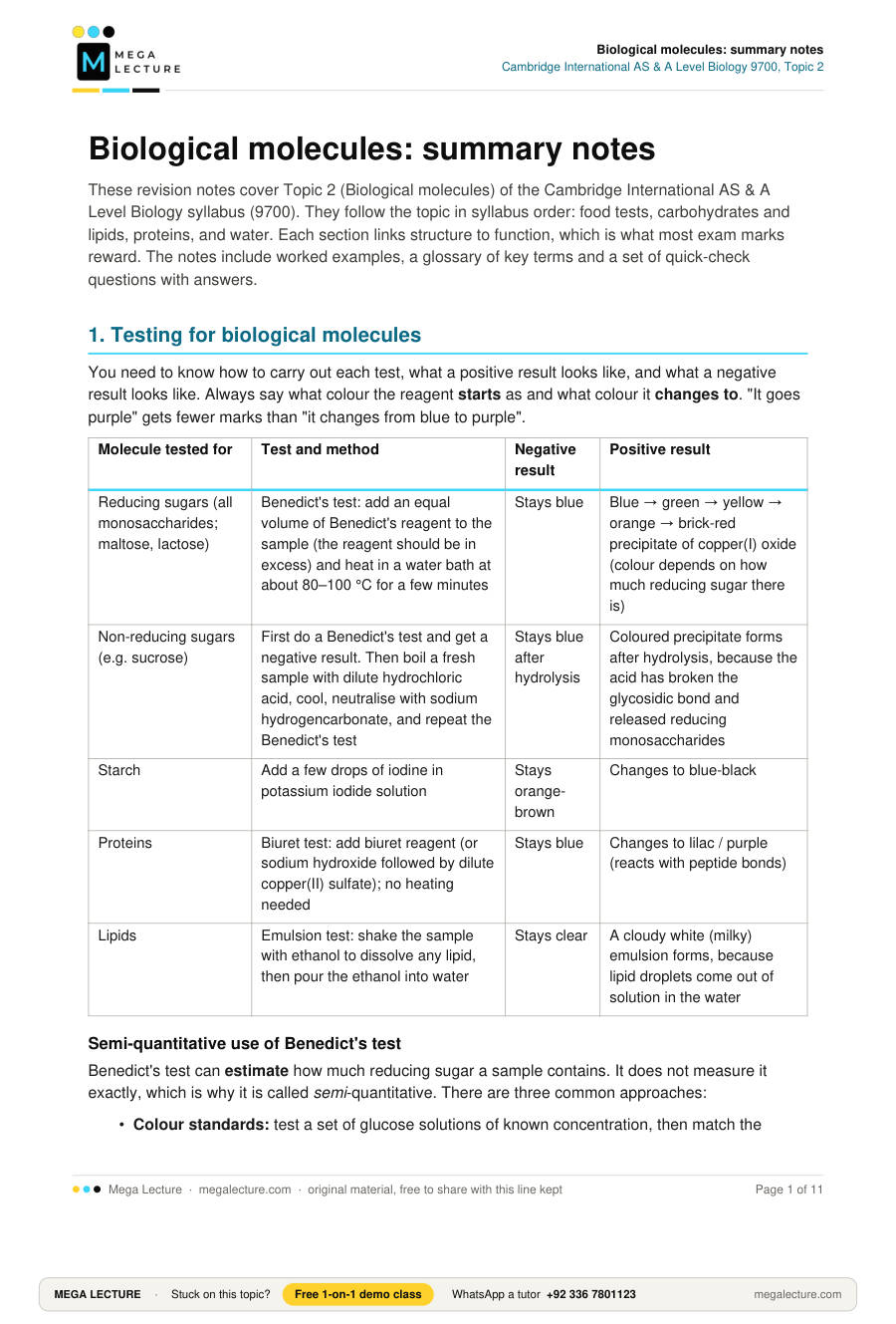
1. Testing for biological molecules
You need to know how to carry out each test, what a positive result looks like, and what a negative result looks like. Always say what colour the reagent starts as and what colour it changes to. "It goes purple" gets fewer marks than "it changes from blue to purple".
| Molecule tested for | Test and method | Negative result | Positive result |
|---|---|---|---|
| Reducing sugars (all monosaccharides; maltose, lactose) | Benedict's test: add an equal volume of Benedict's reagent to the sample (the reagent should be in excess) and heat in a water bath at about 80–100 °C for a few minutes | Stays blue | Blue → green → yellow → orange → brick-red precipitate of copper(I) oxide (colour depends on how much reducing sugar there is) |
| Non-reducing sugars (e.g. sucrose) | First do a Benedict's test and get a negative result. Then boil a fresh sample with dilute hydrochloric acid, cool, neutralise with sodium hydrogencarbonate, and repeat the Benedict's test | Stays blue after hydrolysis | Coloured precipitate forms after hydrolysis, because the acid has broken the glycosidic bond and released reducing monosaccharides |
| Starch | Add a few drops of iodine in potassium iodide solution | Stays orange-brown | Changes to blue-black |
| Proteins | Biuret test: add biuret reagent (or sodium hydroxide followed by dilute copper(II) sulfate); no heating needed | Stays blue | Changes to lilac / purple (reacts with peptide bonds) |
| Lipids | Emulsion test: shake the sample with ethanol to dissolve any lipid, then pour the ethanol into water | Stays clear | A cloudy white (milky) emulsion forms, because lipid droplets come out of solution in the water |
Semi-quantitative use of Benedict's test
Benedict's test can estimate how much reducing sugar a sample contains. It does not measure it exactly, which is why it is called semi-quantitative. There are three common approaches:
- Colour standards: test a set of glucose solutions of known concentration, then match the colour of the unknown to the nearest standard.
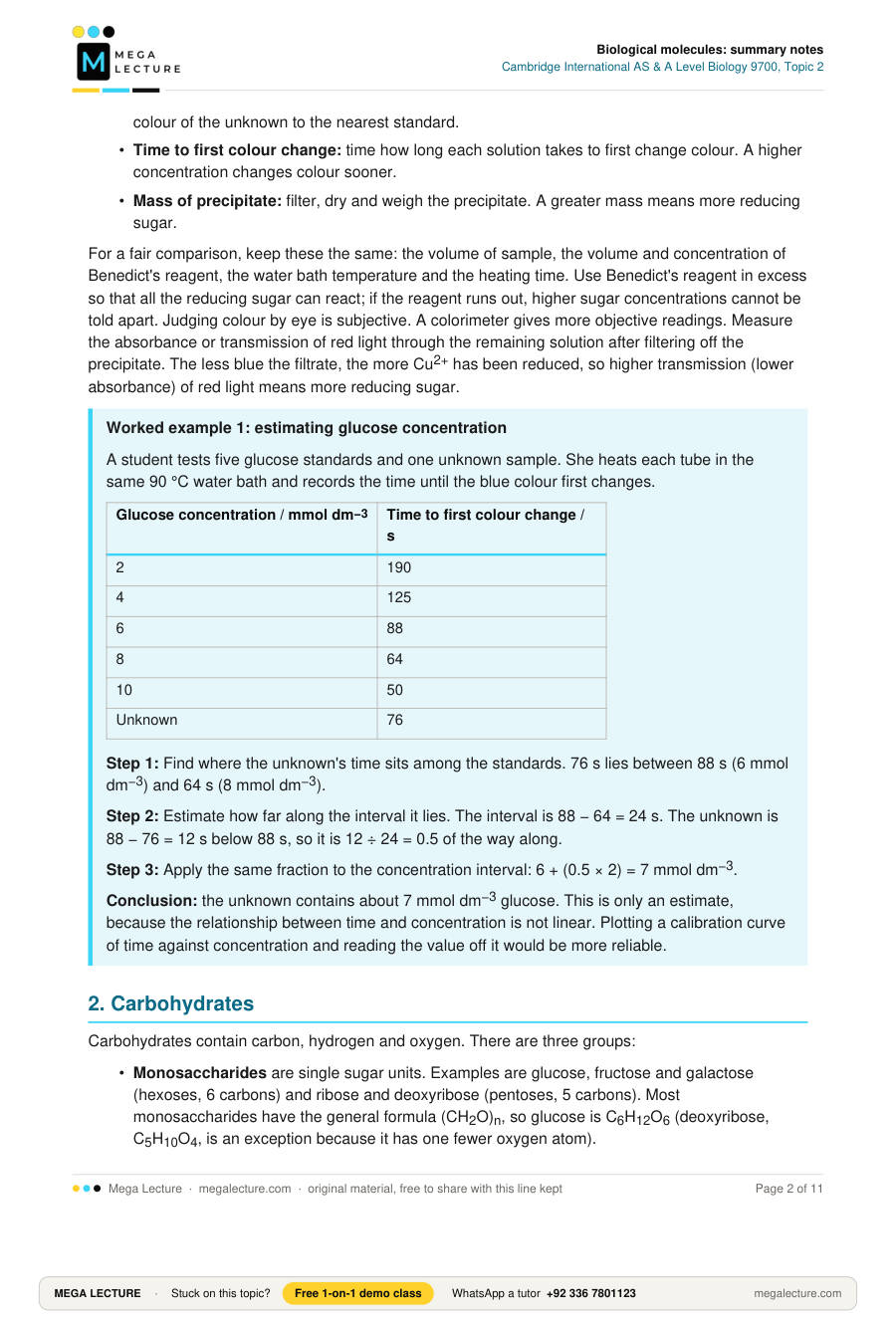
- Time to first colour change: time how long each solution takes to first change colour. A higher concentration changes colour sooner.
- Mass of precipitate: filter, dry and weigh the precipitate. A greater mass means more reducing sugar.
For a fair comparison, keep these the same: the volume of sample, the volume and concentration of Benedict's reagent, the water bath temperature and the heating time. Use Benedict's reagent in excess so that all the reducing sugar can react; if the reagent runs out, higher sugar concentrations cannot be told apart. Judging colour by eye is subjective. A colorimeter gives more objective readings. Measure the absorbance or transmission of red light through the remaining solution after filtering off the precipitate. The less blue the filtrate, the more Cu2+ has been reduced, so higher transmission (lower absorbance) of red light means more reducing sugar.
Worked example 1: estimating glucose concentration
A student tests five glucose standards and one unknown sample. She heats each tube in the same 90 °C water bath and records the time until the blue colour first changes.
| Glucose concentration / mmol dm−3 | Time to first colour change / s |
|---|---|
| 2 | 190 |
| 4 | 125 |
| 6 | 88 |
| 8 | 64 |
| 10 | 50 |
| Unknown | 76 |
Step 1: Find where the unknown's time sits among the standards. 76 s lies between 88 s (6 mmol dm−3) and 64 s (8 mmol dm−3).
Step 2: Estimate how far along the interval it lies. The interval is 88 − 64 = 24 s. The unknown is 88 − 76 = 12 s below 88 s, so it is 12 ÷ 24 = 0.5 of the way along.
Step 3: Apply the same fraction to the concentration interval: 6 + (0.5 × 2) = 7 mmol dm−3.
Conclusion: the unknown contains about 7 mmol dm−3 glucose. This is only an estimate, because the relationship between time and concentration is not linear. Plotting a calibration curve of time against concentration and reading the value off it would be more reliable.
2. Carbohydrates
Carbohydrates contain carbon, hydrogen and oxygen. There are three groups:
- Monosaccharides are single sugar units. Examples are glucose, fructose and galactose (hexoses, 6 carbons) and ribose and deoxyribose (pentoses, 5 carbons). Most monosaccharides have the general formula (CH2O)n, so glucose is C6H12O6 (deoxyribose, C5H10O4, is an exception because it has one fewer oxygen atom).
- Disaccharides are two monosaccharides joined together. Maltose is glucose + glucose, sucrose is glucose + fructose, and lactose is glucose + galactose.
- Polysaccharides are many monosaccharides joined together. Examples are starch, glycogen and cellulose.
Alpha and beta glucose
Glucose forms a ring of five carbon atoms and one oxygen atom. Carbon 6 sits outside the ring. There are two isomers, and they differ only at carbon 1:
| Feature | α-glucose | β-glucose |
|---|---|---|
| –OH group on carbon 1 (ring drawn in the standard way) | Below the ring, on the same side as the –OH on carbon 4 | Above the ring, on the opposite side to the –OH on carbon 4 |
| Polymers formed | Starch (amylose, amylopectin) and glycogen | Cellulose |
Glycosidic bonds: condensation and hydrolysis
Learn these definitions
Condensation reaction: a reaction in which two molecules are joined by a covalent bond, with the removal of a molecule of water.
Hydrolysis reaction: a reaction in which a covalent bond is broken by the addition of a molecule of water.
Glycosidic bond: the covalent C–O–C bond that links two monosaccharides, formed by a condensation reaction.
When two α-glucose molecules join between carbon 1 of one and carbon 4 of the other, they form an α-1,4 glycosidic bond and the disaccharide maltose:
C6H12O6 + C6H12O6 → C12H22O11 + H2O
Hydrolysis reverses this reaction. In the body, enzymes such as maltase catalyse it. In the laboratory, boiling with dilute acid does the same job.
Worked example 2: masses and water in condensation
(a) Calculate the relative molecular mass of maltose. (Ar: C = 12, H = 1, O = 16)
Step 1: Find Mr of glucose, C6H12O6: (6 × 12) + (12 × 1) + (6 × 16) = 72 + 12 + 96 = 180.
Step 2: Two glucose molecules have a total Mr of 2 × 180 = 360.
Step 3: Condensation removes one water molecule (Mr = 18): 360 − 18 = 342. Check: C12H22O11 = 144 + 22 + 176 = 342, which agrees.
(b) An unbranched amylose chain contains 600 glucose units. How many water molecules were released when it formed?
Step 1: Each glycosidic bond releases one water molecule.
Step 2: A straight chain of n units has n − 1 bonds between them, so 600 − 1 = 599 water molecules were released.
Step 3: Hydrolysing the chain completely would therefore use 599 water molecules.
Polysaccharides: structure and function
| Feature | Amylose | Amylopectin | Glycogen | Cellulose |
|---|---|---|---|---|
| Monomer | α-glucose | α-glucose | α-glucose | β-glucose |
| Glycosidic bonds | 1,4 only | 1,4 plus 1,6 at branch points | 1,4 plus 1,6 at branch points | 1,4 only; each glucose is rotated 180° relative to the next |
| Shape | Unbranched chain that coils into a helix, held by hydrogen bonds | Branched (a branch about every 20–30 glucose units) | Very highly branched (a branch about every 8–12 glucose units), with shorter chains and more branches than amylopectin | Long straight chains lying side by side |
| Where found | Plants (part of starch) | Plants (part of starch) | Animals and fungi (liver, muscle) | Plant cell walls |
| Function | Energy store | Energy store | Energy store | Structural support |
How structure fits function in storage polysaccharides:
- They are large and insoluble, so they do not affect the water potential of the cell and cannot diffuse out of it.
- The amylose helix is compact, so a lot of glucose fits in a small space.
- Branching gives amylopectin, and even more so glycogen, many free chain ends. Enzymes can hydrolyse many ends at once and release glucose quickly. This suits animals, which have high and changing respiratory demands.
How structure fits function in cellulose:
- Neighbouring β-glucose units are rotated 180° from each other, so the chain stays straight rather than coiling.
- –OH groups stick out on both sides of each chain, which lets many hydrogen bonds form between parallel chains.
- Many parallel cellulose chains, held together by hydrogen bonds, form microfibrils, and microfibrils group into fibres. Together these give very high tensile strength, so the cell wall can resist the pressure of a turgid cell without bursting.
- Most animals lack an enzyme that can hydrolyse β-1,4 bonds, so they cannot digest cellulose.
3. Lipids
Triglycerides
A triglyceride forms when one glycerol molecule joins to three fatty acids in three condensation reactions. These reactions form three ester bonds and release three water molecules. Each ester bond forms between an –OH group of glycerol and the –COOH (carboxyl) group of a fatty acid.
Ester bond: the covalent bond formed by a condensation reaction between a hydroxyl (–OH) group of glycerol and the carboxyl (–COOH) group of a fatty acid.
| Feature | Saturated fatty acid | Unsaturated fatty acid |
|---|---|---|
| Carbon–carbon bonds in the hydrocarbon chain | Only single C–C bonds | One (mono-) or more (poly-unsaturated) C=C double bonds |
| Shape of chain | Straight, so chains pack closely | Has a kink at each (cis) C=C double bond, so chains cannot pack closely |
| Triglycerides rich in these fatty acids, at room temperature | Solid (fats, mostly from animals) | Liquid (oils, mostly from plants) |
Roles of triglycerides, linked to structure:
- Energy storage: the fatty acid chains contain many C–H bonds. Oxidising a gram of triglyceride releases roughly twice as much energy as a gram of carbohydrate.
- Insoluble in water: they are non-polar, so they do not affect water potential.
- Thermal insulation: fat under the skin (for example blubber in whales) reduces heat loss.
- Buoyancy: fat is less dense than water, which helps aquatic mammals float.
- Protection: fat around organs such as the kidneys cushions them.
- Metabolic water: oxidising triglycerides produces a lot of water, which matters for desert animals.
Phospholipids
In a phospholipid, one of the three fatty acids of a triglyceride is replaced by a phosphate group. This gives the molecule two different ends:
- A hydrophilic head. The phosphate group is charged, so it is attracted to water.
- Two hydrophobic tails. The fatty acid chains are non-polar, so they are repelled by water.
In water, phospholipids arrange themselves into a bilayer. The heads face the watery fluid on each side and the tails point inwards, away from water. This bilayer is the basis of every cell membrane. Its hydrophobic core blocks water-soluble substances such as ions and glucose, so the membrane can control what enters and leaves the cell. Unsaturated tails, with their kinks, make the membrane more fluid.
4. Proteins
Amino acids and peptide bonds
Every amino acid has a central carbon atom bonded to four groups:
- a hydrogen atom
- an amine group (–NH2)
- a carboxyl group (–COOH)
- a variable R group (side chain)
About 20 different R groups are found in proteins. Some are small (glycine's R group is just –H), some are hydrophobic, some are polar, some are charged, and some contain sulfur (cysteine and methionine). Cysteine's –SH group can form disulfide bonds.
Peptide bond: the covalent C–N bond formed by a condensation reaction between the carboxyl group of one amino acid and the amine group of another, with the release of one molecule of water.
Two amino acids make a dipeptide. Many amino acids make a polypeptide. Hydrolysis of peptide bonds, for example by protease enzymes, reverses the reaction.
Levels of protein structure
| Level | What it is | Bonds that hold it |
|---|---|---|
| Primary | The sequence of amino acids in a polypeptide chain | Peptide bonds |
| Secondary | Regular folding or coiling of the chain into an α-helix or a β-pleated sheet | Hydrogen bonds between the –C=O of one peptide bond and the –NH of another along the backbone |
| Tertiary | Further folding of the whole chain into a specific 3D shape | Interactions between R groups: hydrogen bonds, ionic bonds, disulfide bonds and hydrophobic interactions |
| Quaternary | Two or more polypeptide chains joined into one functional protein (sometimes with a non-protein prosthetic group) | The same bonds as tertiary, but between different chains |
Bonds that stabilise tertiary and quaternary structure. Disulfide bonds are the strongest because they are covalent. The others are weaker and are broken by high temperature and by changes in pH.
- Disulfide bonds: covalent S–S bonds between the R groups of two cysteine residues. They are strong and not broken by heat alone.
- Ionic bonds: form between oppositely charged R groups (–NH3+ and –COO−). Changes in pH alter these charges, and high temperature also breaks them.
- Hydrogen bonds: form between polar R groups. Each one is weak, but there are many of them. Heat breaks them easily, and pH changes can also disrupt them.
- Hydrophobic interactions: non-polar R groups cluster together in the centre of the molecule, away from water.
The primary structure decides where each R group sits, so it decides which bonds can form and therefore the final shape. Changing one amino acid can change the shape and function of the whole protein.
Globular and fibrous proteins
| Feature | Globular (e.g. haemoglobin) | Fibrous (e.g. collagen) |
|---|---|---|
| Shape | Compact, roughly spherical | Long strands |
| Solubility in water | Soluble | Insoluble |
| Amino acid sequence | Irregular | Often a repeating pattern |
| Role | Physiological or metabolic (enzymes, transport, hormones, antibodies) | Structural (tendons, skin, bone, hair) |
Haemoglobin: a globular protein
- Its quaternary structure has four polypeptide chains: two α-globin chains and two β-globin chains.
- Each chain holds one haem group, a non-protein prosthetic group that contains an iron ion (Fe2+). Each haem can bind one O2 molecule, so one haemoglobin molecule carries up to four O2 molecules.
- Hydrophobic R groups point into the centre of the molecule and hydrophilic R groups point outwards. This makes haemoglobin soluble in the cytoplasm of red blood cells.
- (Extension: links to the inheritance topic.) In sickle cell anaemia, one amino acid in the β chain changes: glutamic acid (charged, hydrophilic R group) is replaced by valine (non-polar, hydrophobic R group). The haemoglobin becomes less soluble at low oxygen concentration and forms long fibres that distort the red blood cell.
Collagen: a fibrous protein
- Each collagen molecule is made of three polypeptide chains, each a tight helix. The three chains wind around each other to form a triple helix.
- Glycine, the smallest amino acid, is every third amino acid. Its small size lets the three chains pack very closely together.
- Many hydrogen bonds hold the three chains together.
- Covalent cross-links form between neighbouring collagen molecules. The molecules are staggered, so their ends do not line up and there is no weak point.
- Collagen molecules group into fibrils, and fibrils group into fibres. This gives very high tensile strength, which suits tendons, skin, bone and artery walls. Because collagen is insoluble, it stays in place in these tissues.
5. Water
A water molecule is polar. Oxygen attracts the shared electrons more strongly than hydrogen does, so the oxygen carries a small negative charge (δ−) and each hydrogen carries a small positive charge (δ+). A hydrogen bond is the weak attraction between the δ+ hydrogen of one water molecule and the δ− oxygen of another. Each bond is weak, but water contains huge numbers of them, and they explain water's properties.
| Property | Why it happens | Importance to living organisms |
|---|---|---|
| Solvent | Polar water molecules cluster around ions and polar molecules and separate them from each other | Chemical reactions in cells take place in solution. Water transports dissolved substances in blood plasma, in xylem and phloem, and removes urea in urine. |
| High specific heat capacity (about 4.2 J g−1 °C−1) | A lot of energy is needed to break hydrogen bonds before the molecules can move faster and the temperature can rise | The temperature of cells, bodies and aquatic habitats stays fairly stable, which protects enzymes from large temperature swings |
| High latent heat of vaporisation | A lot of energy is needed to break hydrogen bonds so that molecules can escape as vapour | Evaporation of a small amount of water removes a lot of heat. Sweating in mammals and transpiration in leaves both provide cooling. |
Worked example 3: specific heat capacity
Calculate the energy needed to raise the temperature of 250 g of water from 20 °C to 35 °C. Specific heat capacity of water = 4.2 J g−1 °C−1.
Step 1: Temperature change: Δθ = 35 − 20 = 15 °C.
Step 2: Energy = mass × specific heat capacity × Δθ = 250 × 4.2 × 15.
Step 3: 250 × 4.2 = 1050, and 1050 × 15 = 15 750 J.
Step 4: Convert to kJ: 15 750 ÷ 1000 = 15.75 kJ (about 16 kJ).
Evaporating just 7 g of water at body temperature (about 2.4 kJ g−1) would remove roughly 7 × 2.4 ≈ 17 kJ, which is more than all that heating. This is why sweating cools the body so effectively.
Key terms
| Term | Definition |
|---|---|
| Monomer | A small molecule that can join to other, similar molecules to form a polymer |
| Polymer | A large molecule made of many repeating monomer units joined by covalent bonds |
| Macromolecule | A very large molecule, such as a polysaccharide, protein or nucleic acid |
| Monosaccharide | A single sugar unit; the monomer of carbohydrates (e.g. glucose) |
| Disaccharide | A sugar made of two monosaccharides joined by a glycosidic bond (e.g. maltose) |
| Polysaccharide | A polymer made of many monosaccharides joined by glycosidic bonds (e.g. starch) |
| Reducing sugar | A sugar that can donate electrons to (reduce) the Cu2+ ions in Benedict's reagent, forming a brick-red precipitate of copper(I) oxide |
| Condensation | A reaction that joins two molecules with a covalent bond and removes a water molecule |
| Hydrolysis | A reaction that breaks a covalent bond by adding a water molecule |
| Glycosidic bond | A covalent bond between two monosaccharides |
| Ester bond | A covalent bond between glycerol and a fatty acid |
| Peptide bond | A covalent bond between the carboxyl group of one amino acid and the amine group of another |
| Hydrophilic / hydrophobic | Attracted to water / repelled by water |
| Primary structure | The sequence of amino acids in a polypeptide |
| Prosthetic group | A non-protein component that is permanently bound to a protein and needed for its function (e.g. haem) |
| Disulfide bond | A covalent bond between the sulfur atoms in the R groups of two cysteine residues |
| Tensile strength | The ability to resist being pulled apart |
| Specific heat capacity | The energy needed to raise the temperature of 1 g of a substance by 1 °C |
| Latent heat of vaporisation | The energy needed to change 1 g of a liquid into vapour without changing its temperature |
Quick check
- State the colour change in a positive biuret test. [1]
- Name the monomer of cellulose and the type of bond that links the monomers. [2]
- State the number of water molecules released when one triglyceride is formed, and name the bonds formed. [2]
- Describe how you would test a solution for a non-reducing sugar. [4]
- Calculate the relative molecular mass of a trisaccharide made from three glucose molecules (Mr of glucose = 180, water = 18). [2]
- Outline how a hydrogen bond forms between two water molecules. [3]
- Compare the structure of amylose with the structure of glycogen. [3]
- Explain why glycogen is well suited to its role as an energy store in animals. [3]
- Explain how the structure of a phospholipid lets it form a bilayer in water. [3]
- Explain how the structure of collagen gives it high tensile strength. [4]
- A protein is heated to 60 °C and loses its function. Analysis shows that its primary structure has not changed. Explain these observations. [3]
- Two students test the same unknown solution with Benedict's reagent. One judges the result "orange" and the other "yellow". Suggest how the investigation could be improved to give a more reliable estimate of reducing sugar concentration. [3]
- Describe the difference between α-glucose and β-glucose. [1]
- Explain how the structure of haemoglobin is related to its function. [4]
- Explain why sweating cools the body. [2]
Answers
- Blue → lilac / purple. [1]
- β-glucose [1]; (β-1,4) glycosidic bond [1].
- Three water molecules [1]; ester bonds [1].
- Test first with Benedict's reagent and heat; the result is negative and the solution stays blue [1]. Heat a fresh sample with dilute hydrochloric acid to hydrolyse glycosidic bonds [1]. Cool, then neutralise with sodium hydrogencarbonate [1]. Add Benedict's reagent and heat; a colour change from blue to green/yellow/orange/brick-red shows a non-reducing sugar was present [1].
- 3 × 180 = 540; two glycosidic bonds form, so two water molecules are removed: 540 − (2 × 18) [1] = 504 [1].
- Water is polar: oxygen is δ− and the hydrogens are δ+ [1], because oxygen attracts the shared electrons more strongly [1]. The δ+ hydrogen of one molecule is attracted to the δ− oxygen of a neighbouring molecule [1].
- Both are made of α-glucose linked by 1,4 glycosidic bonds [1]. Amylose is unbranched and coils into a helix, while glycogen is highly branched [1]. Glycogen has 1,6 glycosidic bonds at branch points, but amylose does not [1].
- It is insoluble, so it does not lower water potential or leave the cell [1]. It is compact, so a lot of glucose is stored in a small space [1]. It is highly branched, with many ends for enzymes to hydrolyse, so glucose is released rapidly for respiration [1].
- The phosphate head is charged/polar and hydrophilic, so it faces water [1]. The two fatty acid tails are non-polar and hydrophobic, so they point away from water [1]. Two layers therefore form, with tails facing inwards and heads facing the watery fluid on each side [1].
- Any four from: three polypeptide chains wound into a triple helix [1]; glycine every third amino acid lets the chains pack tightly [1]; many hydrogen bonds between the chains [1]; covalent cross-links between neighbouring molecules [1]; molecules staggered so there is no weak point [1]; molecules grouped into fibrils and then fibres [1].
- Heat breaks weak bonds that hold the 3D (tertiary) shape, such as hydrogen bonds and ionic bonds [1]. The protein unfolds / is denatured, so its specific shape (for example an enzyme's active site) is lost and it cannot function [1]. Peptide bonds are covalent and are not broken by this heating, so the amino acid sequence stays the same [1].
- Any three from: use a colorimeter to measure the colour objectively rather than by eye [1]; prepare a calibration curve from glucose standards of known concentration [1]; control volumes of sample and reagent, water bath temperature and heating time [1]; use Benedict's reagent in excess [1]; repeat the test and calculate a mean [1]; or filter, dry and weigh the precipitate [1].
- In α-glucose the –OH on carbon 1 is below the ring (same side as the –OH on carbon 4); in β-glucose it is above the ring (opposite side) [1].
- Any four from: four polypeptide chains give a quaternary structure [1]; each chain has a haem group containing Fe2+ [1]; each haem binds one O2, so one molecule carries up to four O2 [1]; hydrophobic R groups point inwards and hydrophilic R groups point outwards [1]; so haemoglobin is soluble in the cytoplasm of red blood cells [1].
- Water in sweat evaporates from the skin, and water has a high latent heat of vaporisation [1]. A lot of heat energy is taken from the body to break hydrogen bonds as the water evaporates, so a small amount of evaporation removes a lot of heat [1].
