
A Level · Biology 9700 · Cell structure: summary notes
Cell structure: summary notes
Opens inside this page. Prefer your own PDF app? Open the PDF.
Check yourself
5 quick questions on A Level Biology
Tap an answer to see if you are right, and why. Free, no sign-up.
Start the free test12 of 12 students found these notes useful
Was this note useful?
Full text of Cell structure: summary notes
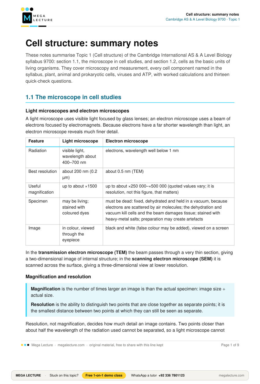
These notes summarise Topic 1 (Cell structure) of the Cambridge International AS & A Level Biology syllabus 9700: section 1.1, the microscope in cell studies, and section 1.2, cells as the basic units of living organisms. They cover microscopy and measurement, every cell component named in the syllabus, plant, animal and prokaryotic cells, viruses and ATP, with worked calculations and thirteen quick-check questions.
1.1 The microscope in cell studies
Light microscopes and electron microscopes
A light microscope uses visible light focused by glass lenses; an electron microscope uses a beam of electrons focused by electromagnets. Because electrons have a far shorter wavelength than light, an electron microscope reveals much finer detail.
| Feature | Light microscope | Electron microscope |
|---|---|---|
| Radiation | visible light, wavelength about 400–700 nm | electrons, wavelength well below 1 nm |
| Best resolution | about 200 nm (0.2 µm) | about 0.5 nm (TEM) |
| Useful magnification | up to about ×1500 | up to about ×250 000–×500 000 (quoted values vary; it is resolution, not this figure, that matters) |
| Specimen | may be living; stained with coloured dyes | must be dead: fixed, dehydrated and held in a vacuum, because electrons are scattered by air molecules; the dehydration and vacuum kill cells and the beam damages tissue; stained with heavy-metal salts; preparation may create artefacts |
| Image | in colour, viewed through the eyepiece | black and white (false colour may be added), viewed on a screen |
In the transmission electron microscope (TEM) the beam passes through a very thin section, giving a two-dimensional image of internal structure; in the scanning electron microscope (SEM) it is scanned across the surface, giving a three-dimensional view at lower resolution.
Magnification and resolution
Magnification is the number of times larger an image is than the actual specimen: image size ÷ actual size.
Resolution is the ability to distinguish two points that are close together as separate points; it is the smallest distance between two points at which they can still be seen as separate.
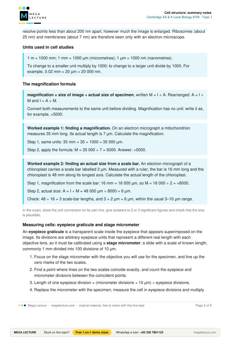
Resolution, not magnification, decides how much detail an image contains. Two points closer than about half the wavelength of the radiation used cannot be separated, so a light microscope cannot resolve points less than about 200 nm apart, however much the image is enlarged. Ribosomes (about 25 nm) and membranes (about 7 nm) are therefore seen only with an electron microscope.
Units used in cell studies
1 m = 1000 mm; 1 mm = 1000 µm (micrometres); 1 µm = 1000 nm (nanometres).
To change to a smaller unit multiply by 1000; to change to a larger unit divide by 1000. For example, 0.02 mm = 20 µm = 20 000 nm.
The magnification formula
magnification = size of image ÷ actual size of specimen, written M = I ÷ A. Rearranged: A = I ÷ M and I = A × M.
Convert both measurements to the same unit before dividing. Magnification has no unit: write it as, for example, ×5000.
Worked example 1: finding a magnification. On an electron micrograph a mitochondrion measures 35 mm long. Its actual length is 7 µm. Calculate the magnification.
Step 1, same units: 35 mm = 35 × 1000 = 35 000 µm.
Step 2, apply the formula: M = 35 000 ÷ 7 = 5000. Answer: ×5000.
Worked example 2: finding an actual size from a scale bar. An electron micrograph of a chloroplast carries a scale bar labelled 2 µm. Measured with a ruler, the bar is 16 mm long and the chloroplast is 48 mm along its longest axis. Calculate the actual length of the chloroplast.
Step 1, magnification from the scale bar: 16 mm = 16 000 µm, so M = 16 000 ÷ 2 = ×8000.
Step 2, actual size: A = I ÷ M = 48 000 µm ÷ 8000 = 6 µm.
Check: 48 ÷ 16 = 3 scale-bar lengths, and 3 × 2 µm = 6 µm, within the usual 3–10 µm range.
In the exam, show the unit conversion on its own line, give answers to 2 or 3 significant figures and check that the size is plausible.
Measuring cells: eyepiece graticule and stage micrometer
An eyepiece graticule is a transparent scale inside the eyepiece that appears superimposed on the image. Its divisions are arbitrary eyepiece units that represent a different real length with each objective lens, so it must be calibrated using a stage micrometer: a slide with a scale of known length, commonly 1 mm divided into 100 divisions of 10 µm.
- Focus on the stage micrometer with the objective you will use for the specimen, and line up the zero marks of the two scales.
- Find a point where lines on the two scales coincide exactly, and count the eyepiece and micrometer divisions between the coincident points.
- Length of one eyepiece division = (micrometer divisions × 10 µm) ÷ eyepiece divisions.
- Replace the micrometer with the specimen, measure the cell in eyepiece divisions and multiply by the calibration value. Recalibrate whenever the objective is changed.
Worked example 3: calibrating and measuring. Using the ×40 objective, a student finds that 40 eyepiece divisions coincide with 10 stage micrometer divisions of 10 µm each.
10 micrometer divisions = 100 µm, so one eyepiece division = 100 ÷ 40 = 2.5 µm.
With the same objective, a palisade mesophyll cell spans 28 eyepiece divisions. Its length = 28 × 2.5 = 70 µm.
Check: with the ×10 objective the same graticule gives about 10 µm per division, four times as much, so the calibration value must be found again for each objective.
Temporary preparations and drawing rules
To make a temporary mount, place a thin piece of tissue (one or a few cells thick, so that light can pass through it) on a slide, add a drop of water or stain, lower a coverslip slowly at an angle with a mounting needle so that no air bubbles are trapped, and blot any excess liquid with filter paper. Iodine in potassium iodide solution stains starch blue-black; methylene blue stains the nuclei of animal cells.
Drawings from slides and photomicrographs are marked for accuracy, not artistry:
- Use a sharp HB pencil; draw single, clear, continuous lines with no sketching, shading or colouring; make the drawing large, keep the proportions correct and draw only what you can actually see.
- Draw label lines with a ruler, ending exactly on the structure, with no arrowheads and no crossing; write labels horizontally outside the drawing.
- Give a title and state the magnification, or add a scale bar.
- A low-power plan shows only the outlines of tissues, with no individual cells; a high-power drawing shows a few cells in detail, with walls drawn as double lines.
1.2 Cells as the basic units of living organisms
All living organisms are made of cells, the smallest units that can carry out all the processes of life. Eukaryotic cells (animals, plants, fungi and protoctists) have a nucleus and other membrane-bound organelles, which keep incompatible reactions apart.
Structure and function of eukaryotic cell components
| Component | Structure | Function |
|---|---|---|
| Cell surface membrane | Phospholipid bilayer with proteins, about 7 nm thick. | Partially permeable barrier controlling what enters and leaves the cell; carries receptors and cell-recognition glycoproteins. |
| Nucleus | Largest organelle, typically about 10 µm across; contains chromatin (DNA bound to histone proteins), which condenses into chromosomes during nuclear division. | Stores genetic information and controls the cell's activities; site of DNA replication and transcription. |
| Nuclear envelope | Two membranes with nuclear pores; the outer membrane is continuous with the rough ER. | Separates DNA from the cytoplasm; pores let mRNA and ribosomal subunits out and proteins in. |
| Nucleolus | Dense region inside the nucleus, with no membrane. | Makes ribosomal RNA and assembles ribosomal subunits. |
| Rough endoplasmic reticulum (RER) | Flattened membrane sacs (cisternae) with 80S ribosomes on the outer surface. | Proteins made by the ribosomes enter the cisternae and travel in vesicles to the Golgi body; makes proteins for secretion and for membranes. |
| Smooth endoplasmic reticulum (SER) | Similar membrane system, more tubular, with no ribosomes. | Makes lipids and steroids (e.g. cholesterol, reproductive hormones); detoxifies drugs in liver cells. |
| Golgi body (Golgi apparatus) | Stack of curved, flattened sacs with vesicles pinching off the edges. | Modifies proteins (e.g. adds carbohydrate to make glycoproteins) and packages them into vesicles for secretion by exocytosis; makes lysosomes; in plant cells makes the non-cellulose cell wall polysaccharides (pectins, hemicelluloses). |
| Mitochondrion | About 1 µm wide; envelope of two membranes, the inner folded into cristae; the matrix contains enzymes, small circular DNA and 70S ribosomes. | Site of aerobic respiration, producing most of the cell's ATP; the cristae give a large surface area for the enzymes and proteins of the final stages. |
| Ribosomes | Particles of rRNA and protein with a large and a small subunit; 80S (about 25 nm) free in the cytoplasm or on the RER; 70S (about 20 nm) in mitochondria, chloroplasts and prokaryotes. | Site of protein synthesis (translation). |
| Lysosomes | Spherical sacs bounded by a single membrane, formed by the Golgi body; contain hydrolytic enzymes (proteases, lipases, nucleases). | Digest worn-out organelles and material taken in by phagocytosis, such as bacteria; can destroy the whole cell (autolysis). |
| Centrioles and centrosome | The centrosome is a region of cytoplasm next to the nucleus of an animal cell containing a pair of centrioles: hollow cylinders at right angles to each other, each made of nine microtubule triplets. | The centrosome is the microtubule-organising centre (MTOC) from which the spindle microtubules grow during mitosis and meiosis; the centrioles themselves are not needed for spindle formation (plant cells have no centrioles but still form a spindle from an MTOC). Centrioles act as basal bodies, the MTOCs that assemble the microtubules of cilia and flagella. |
| Microtubules | Hollow tubes about 25 nm across, made of the protein tubulin. | Cytoskeleton giving shape and support; tracks for moving vesicles and organelles; make up spindle fibres, centrioles and cilia. |
| Cilia | Hair-like projections a few µm long, with nine microtubule doublets around a central pair (9 + 2), covered by cell surface membrane. | Beat, using ATP, to move fluid across a surface (e.g. mucus in the airways) or to move the cell; flagella are similar but longer. |
| Microvilli | Finger-like folds of the cell surface membrane, about 1 µm long. | Increase surface area for absorption, e.g. in the epithelium of the small intestine. |
| Chloroplast | 3–10 µm long; envelope of two membranes; thylakoid membranes stacked into grana within the stroma, which contains starch grains, small circular DNA and 70S ribosomes; chlorophyll is in the thylakoids. | Site of photosynthesis: ATP is made on the thylakoids in the light-dependent stage and used in the stroma to make sugars. |
| Cell wall | Rigid layer of cellulose microfibrils outside the plant cell surface membrane; adjacent cells are cemented by the middle lamella; fully permeable. | Strength and fixed shape; stops the cell bursting when it absorbs water, so it becomes turgid and supports the plant. |
| Plasmodesmata | Strands of cytoplasm passing through pores in the walls of neighbouring plant cells, lined by cell surface membrane. | Link the cytoplasm of adjacent cells so that substances pass directly between them. |
| Large permanent vacuole and tonoplast | Large central sac of a mature plant cell, bounded by the tonoplast membrane and containing cell sap (water, sugars, ions, amino acids, pigments). | Takes in water to make the cell turgid; stores substances; the tonoplast controls exchange with the cytoplasm. |
Recognising organelles on electron micrographs
- Nucleus: the largest body in the cell, bounded by a double envelope with pores, with dark patches of chromatin and a darker, rounded nucleolus.
- Mitochondrion: oval or sausage-shaped, about 1 µm wide, with two membranes, the inner one folded inwards as cristae.
- Chloroplast: larger (3–10 µm), with two membranes and dark stacks of discs (grana) joined by membranes, often with pale starch grains.
- Rough ER: parallel, flattened membranes dotted with ribosomes; smooth ER: similar but without ribosomes and more tubular.
- Golgi body: a stack of curved, flattened sacs with small vesicles budding from the ends.
- Lysosomes and vesicles: small round bodies bounded by a single membrane.
- Cell wall: a thick, even layer outside the membrane; the middle lamella shows as a line between the walls of neighbouring cells, and plasmodesmata as gaps crossing the wall.
- Vacuole: a large, pale, uniform area filling most of a mature plant cell, bounded by the tonoplast.
An image that shows ribosomes, or membranes as lines, must be an electron micrograph: a two-dimensional section showing internal structure is from a TEM, and a three-dimensional surface view is from an SEM. A light micrograph is in colour and shows the nucleus, cell wall, chloroplasts and vacuole but cannot resolve membranes or ribosomes.
Typical plant and animal cells compared
| Feature | Animal cell | Plant cell |
|---|---|---|
| Cell wall | absent | present, made of cellulose |
| Chloroplasts | absent | present in photosynthesising cells |
| Vacuole | small temporary vacuoles, if any | one large permanent central vacuole bounded by a tonoplast |
| Centrioles | present, in the centrosome | absent (a spindle still forms from a microtubule-organising centre) |
| Lysosomes | present | not usually shown; the large vacuole contains hydrolytic enzymes and carries out the same role |
| Plasmodesmata | absent | present |
| Storage carbohydrate | glycogen granules | starch grains |
| Cilia and microvilli | present on some cells | absent from typical plant cells |
| Shape | irregular, can change | regular, fixed by the wall |
Both have a cell surface membrane, a nucleus with nuclear envelope and nucleolus, rough and smooth ER, a Golgi body, mitochondria, 80S ribosomes and a microtubule cytoskeleton.
ATP: the universal energy currency
ATP (adenosine triphosphate) is a nucleotide made of adenine, ribose and three phosphate groups. It is produced in mitochondria during aerobic respiration and in chloroplasts during the light-dependent stage of photosynthesis. Hydrolysis of the outermost phosphate group (ATP + H2O → ADP + Pi) releases a small, immediately usable amount of energy, and the ADP is then rebuilt into ATP. Every cell of every organism uses ATP as its immediate energy source, hence the universal energy currency. It drives active transport, the synthesis of large molecules such as proteins, and movement such as muscle contraction and the beating of cilia.
Prokaryotic cells
Bacteria are prokaryotes: unicellular, generally 1–5 µm in diameter, with a cell surface membrane but no organelles surrounded by a double membrane (no nucleus, mitochondria or chloroplasts) and no ER, Golgi body or lysosomes. Some have a slimy capsule outside the wall, pili for attachment and one or more flagella for movement.
| Feature | Prokaryotic cell (typical bacterium) | Eukaryotic cell (plant or animal) |
|---|---|---|
| Typical size | 1–5 µm | 10–100 µm |
| Nucleus | absent; DNA lies in the nucleoid region of the cytoplasm | present, bounded by a nuclear envelope with pores |
| DNA | one circular molecule, not associated with histones; small extra rings called plasmids are common | linear molecules combined with histone proteins to form chromosomes |
| Ribosomes | 70S only | 80S in the cytoplasm and on the RER (70S inside mitochondria and chloroplasts) |
| Organelles with a double membrane | none | nucleus, mitochondria and, in plants, chloroplasts |
| ER, Golgi body, lysosomes | absent | present (lysosomes mainly in animal cells) |
| Cell wall | peptidoglycan (murein) | cellulose in plants; chitin in fungi; none in animals |
| Flagella, if present | thin, solid protein filament that rotates | 9 + 2 microtubule structure covered by membrane, which bends |
Viruses
Viruses are not cells. A virus particle is a non-cellular structure, 20–300 nm across, consisting of a core of nucleic acid, either DNA or RNA but never both, surrounded by a protein coat called the capsid, built from repeating protein subunits. Some, such as HIV and influenza virus, also have an outer envelope of phospholipids taken from the host cell membrane. Viruses have no cytoplasm, ribosomes or metabolism of their own, so they reproduce only inside a living host cell, using the host's enzymes and ribosomes.
Quick check
- Define the term resolution. [1]
- State two advantages of a light microscope over an electron microscope, and two advantages of an electron microscope over a light microscope. [4]
- Calculate (a) 0.35 mm in µm, (b) 2.5 µm in nm, (c) 800 nm in µm. [3]
- A red blood cell with an actual diameter of 7.5 µm is drawn 30 mm wide. Calculate the magnification of the drawing. [2]
- An electron micrograph of a bacterium has a magnification of ×20 000. The bacterium measures 60 mm long on the micrograph. Calculate its actual length in µm. [2]
- On an electron micrograph a scale bar labelled 1 µm is 20 mm long. A mitochondrion on the same micrograph is 45 mm long. Calculate its actual length. [2]
- A student calibrated an eyepiece graticule using the ×10 objective, then measured a cell using the ×40 objective and the same calibration value. Explain why the length obtained is wrong, and state whether it is too large or too small. [3]
- Using the ×40 objective, 25 eyepiece graticule divisions line up with 5 stage micrometer divisions of 10 µm each. A cell measures 18 eyepiece divisions. Calculate the length of the cell. [3]
- Identify the cell component that (a) produces most of the cell's ATP, (b) modifies proteins and packages them into vesicles, (c) contains hydrolytic enzymes, (d) makes ribosomal RNA. [4]
- Cells of the pancreas that secrete digestive enzymes contain many mitochondria, a large amount of rough ER and several Golgi bodies. Explain how each of these is involved in secretion. [3]
- State three ways in which the structure of a bacterial cell differs from that of a plant cell. [3]
- Describe the structure of a virus particle. [3]
- Outline the role of ATP in cells. [3]
Answers
- The ability to distinguish two points that are close together as separate points, or the minimum distance between two points at which they can still be seen as separate.
- Light microscope, any two: living specimens can be observed; colour is seen; cheap, portable and quick to use. Electron microscope, any two: far higher resolution (about 0.5 nm compared with 200 nm); far higher useful magnification; shows ribosomes and membranes.
- (a) 350 µm; (b) 2500 nm; (c) 0.8 µm.
- 30 mm = 30 000 µm (1); magnification = 30 000 ÷ 7.5 = ×4000 (1).
- 60 mm = 60 000 µm (1); actual length = 60 000 ÷ 20 000 = 3 µm (1). The answer must carry the unit µm.
- Magnification from the scale bar: 20 mm = 20 000 µm, so M = 20 000 ÷ 1 = ×20 000 (1); actual length = 45 000 µm ÷ 20 000 = 2.25 µm, accept 2.3 µm (1).
- Graticule divisions are arbitrary units that are given a real value only by calibration against the stage micrometer; changing to the ×40 objective magnifies the specimen four times more but does not change the graticule scale, so each division now represents a smaller real length (about 2.5 µm instead of 10 µm); using the ×10 value therefore gives a length that is too large, about four times the true length.
- 5 stage micrometer divisions = 5 × 10 = 50 µm (1); one eyepiece division = 50 ÷ 25 = 2.0 µm (1); cell length = 18 × 2.0 = 36 µm (1).
- (a) mitochondrion; (b) Golgi body; (c) lysosome; (d) nucleolus.
- Ribosomes on the rough ER synthesise the enzyme (a protein), which enters the cisternae and is carried in vesicles to the Golgi body (1); the Golgi body modifies the enzyme and packages it into secretory vesicles, which fuse with the cell surface membrane and release it by exocytosis (1); mitochondria carry out aerobic respiration to supply the ATP needed for protein synthesis and for moving the vesicles and exocytosis (1).
- Any three: cell wall of peptidoglycan rather than cellulose; circular DNA free in the cytoplasm rather than linear DNA bound to histones inside a nucleus; 70S rather than 80S ribosomes in the cytoplasm; no mitochondria, chloroplasts, ER, Golgi body or large permanent vacuole; much smaller (1–5 µm); may have plasmids or a capsule.
- Any three: non-cellular particle (the size, 20–300 nm, is extra detail); core of nucleic acid, either DNA or RNA; surrounded by a protein capsid made of repeating subunits; some have an outer phospholipid envelope derived from the host cell membrane.
- Any three: ATP is the immediate source of energy in every cell (the universal energy currency); made in mitochondria by aerobic respiration and in chloroplasts by photosynthesis; hydrolysis to ADP and phosphate releases a small, manageable amount of energy; this drives active transport, the synthesis of large molecules and movement such as muscle contraction and the beating of cilia.
