
A Level · Biology 9700 · Transport in mammals: summary notes
Transport in mammals: summary notes
Opens inside this page. Prefer your own PDF app? Open the PDF.
Check yourself
5 quick questions on A Level Biology
Tap an answer to see if you are right, and why. Free, no sign-up.
Start the free test12 of 12 students found these notes useful
Was this note useful?
Full text of Transport in mammals: summary notes
These revision notes cover Topic 8, Transport in mammals, of the Cambridge International AS & A Level Biology syllabus (9700). They follow the order of the topic. First come the circulatory system and blood vessels, then blood, tissue fluid and lymph. Next is how oxygen and carbon dioxide are carried: haemoglobin, the oxygen dissociation curve, the Bohr shift, carbonic anhydrase and the chloride shift. The notes end with the heart, the cardiac cycle, and how the heartbeat is started and coordinated. Every step of the worked examples is shown. At the end there is a key terms table and a set of quick-check questions, graded by difficulty, with answers.
1. The mammalian circulatory system
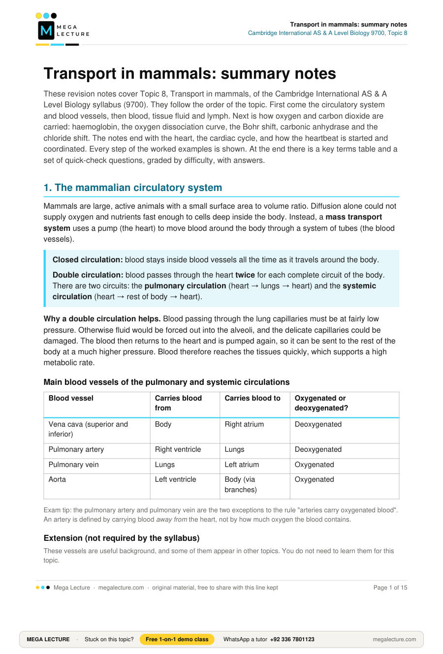
Mammals are large, active animals with a small surface area to volume ratio. Diffusion alone could not supply oxygen and nutrients fast enough to cells deep inside the body. Instead, a mass transport system uses a pump (the heart) to move blood around the body through a system of tubes (the blood vessels).
Closed circulation: blood stays inside blood vessels all the time as it travels around the body.
Double circulation: blood passes through the heart twice for each complete circuit of the body. There are two circuits: the pulmonary circulation (heart → lungs → heart) and the systemic circulation (heart → rest of body → heart).
Why a double circulation helps. Blood passing through the lung capillaries must be at fairly low pressure. Otherwise fluid would be forced out into the alveoli, and the delicate capillaries could be damaged. The blood then returns to the heart and is pumped again, so it can be sent to the rest of the body at a much higher pressure. Blood therefore reaches the tissues quickly, which supports a high metabolic rate.
Main blood vessels of the pulmonary and systemic circulations
| Blood vessel | Carries blood from | Carries blood to | Oxygenated or deoxygenated? |
|---|---|---|---|
| Vena cava (superior and inferior) | Body | Right atrium | Deoxygenated |
| Pulmonary artery | Right ventricle | Lungs | Deoxygenated |
| Pulmonary vein | Lungs | Left atrium | Oxygenated |
| Aorta | Left ventricle | Body (via branches) | Oxygenated |
Exam tip: the pulmonary artery and pulmonary vein are the two exceptions to the rule "arteries carry oxygenated blood". An artery is defined by carrying blood away from the heart, not by how much oxygen the blood contains.
Extension (not required by the syllabus)
These vessels are useful background, and some of them appear in other topics. You do not need to learn them for this topic.
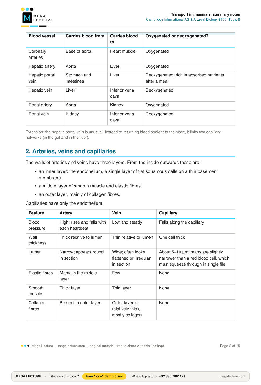
| Blood vessel | Carries blood from | Carries blood to | Oxygenated or deoxygenated? |
|---|---|---|---|
| Coronary arteries | Base of aorta | Heart muscle | Oxygenated |
| Hepatic artery | Aorta | Liver | Oxygenated |
| Hepatic portal vein | Stomach and intestines | Liver | Deoxygenated; rich in absorbed nutrients after a meal |
| Hepatic vein | Liver | Inferior vena cava | Deoxygenated |
| Renal artery | Aorta | Kidney | Oxygenated |
| Renal vein | Kidney | Inferior vena cava | Deoxygenated |
Extension: the hepatic portal vein is unusual. Instead of returning blood straight to the heart, it links two capillary networks (in the gut and in the liver).
2. Arteries, veins and capillaries
The walls of arteries and veins have three layers. From the inside outwards these are:
- an inner layer: the endothelium, a single layer of flat squamous cells on a thin basement membrane
- a middle layer of smooth muscle and elastic fibres
- an outer layer, mainly of collagen fibres.
Capillaries have only the endothelium.
| Feature | Artery | Vein | Capillary |
|---|---|---|---|
| Blood pressure | High; rises and falls with each heartbeat | Low and steady | Falls along the capillary |
| Wall thickness | Thick relative to lumen | Thin relative to lumen | One cell thick |
| Lumen | Narrow; appears round in section | Wide; often looks flattened or irregular in section | About 5–10 µm; many are slightly narrower than a red blood cell, which must squeeze through in single file |
| Elastic fibres | Many, in the middle layer | Few | None |
| Smooth muscle | Thick layer | Thin layer | None |
| Collagen fibres | Present in outer layer | Outer layer is relatively thick, mostly collagen | None |
| Valves | None (except at the base of the aorta and pulmonary artery) | Present (semilunar-shaped pocket valves) | None |
| Endothelium | Often looks wavy or folded in section | Smooth | Squamous; small gaps between cells |
Elastic arteries and muscular arteries
- Elastic arteries are the large arteries close to the heart, such as the aorta and the pulmonary artery. Their middle layer has a very high proportion of elastic fibres, so they can stretch and recoil with each surge of blood from the ventricles.
- Muscular arteries are the arteries further from the heart that deliver blood to organs. Their middle layer has a higher proportion of smooth muscle, so they can narrow or widen to control how much blood goes to each organ.
Relating structure to function
- Elastic fibres in arteries stretch as the ventricles force blood in during systole, then recoil during diastole. This keeps blood moving and evens out the pressure, so flow is smoother further from the heart.
- Smooth muscle can contract to narrow the lumen (vasoconstriction) or relax to widen it (vasodilation). This controls how much blood goes to different organs. Arterioles, the small branches of arteries, have the most important role in this.
- Collagen fibres are strong and resist stretching, so they stop vessel walls from bursting or over-stretching.
- Valves in veins stop blood flowing backwards when pressure is low. When skeletal muscles around a vein contract, they squeeze it, and the valves make sure the blood can only move towards the heart.
- The wide lumen of veins gives little resistance to flow. This matters because venous pressure is low.
- Capillary walls are one cell thick, which gives a very short diffusion distance between the blood and tissue cells. There are huge numbers of capillaries, which gives a large surface area. The narrow lumen slows blood flow, which allows more time for exchange. Gaps between the endothelial cells let plasma leak out to form tissue fluid, and let white blood cells squeeze out.
3. Blood: water, plasma and blood cells
Blood is made of plasma (about 55% of blood volume) and cells (red blood cells, white blood cells and platelets). The main component of plasma is water, and water is also the main component of tissue fluid and lymph.
Why water is a good transport medium
- Water is a solvent. Water molecules are polar, so ions (such as Na+, Cl− and HCO3−) and polar molecules (such as glucose, amino acids, urea and some hormones) dissolve in it and are carried in solution.
- Water has a high specific heat capacity. A lot of energy is needed to change its temperature. Blood can therefore carry heat from warm, active organs (such as the liver and muscles) to cooler parts of the body without large changes in its own temperature.
The syllabus limits this to solvent action and high specific heat capacity, so learn these two. It is also true that water is a liquid at body temperature, so it flows and can be pumped, but that is background only.
Blood cells you must recognise in micrographs and drawings
| Cell | Appearance | Function |
|---|---|---|
| Red blood cell (erythrocyte) | Biconcave disc about 7 µm across; no nucleus; stains evenly, often paler in the centre | Carries oxygen (and some carbon dioxide) |
| Neutrophil | Nucleus with several lobes (usually 2–5); granular cytoplasm; larger than a red blood cell | Phagocytosis of bacteria |
| Monocyte | Largest white cell; large kidney-shaped or bean-shaped nucleus | Leaves the blood and becomes a macrophage, a long-lived phagocyte |
| Lymphocyte | Small; large round nucleus filling almost all of the cell, with only a thin rim of cytoplasm | Specific immune response. B-lymphocytes secrete antibodies. T-lymphocytes include T-helper cells, which release cytokines, and T-killer cells, which destroy infected cells. |
When drawing these cells: use clear, single, unbroken lines and no shading. Draw each nucleus in proportion to the cell (for example, the lymphocyte nucleus fills most of the cell). Label with ruled lines that do not cross.
Red blood cells: structure and function
- The biconcave shape gives a large surface area to volume ratio for fast diffusion of oxygen in and out, and a short diffusion distance to the centre of the cell.
- Mature red blood cells have no nucleus, mitochondria or endoplasmic reticulum, which leaves more room for haemoglobin. Because they have no mitochondria, they respire anaerobically, so they do not use up the oxygen they carry.
- They are small and flexible, so they can squeeze through capillaries that are slightly narrower than themselves. This pushes them close to the capillary wall, which shortens the diffusion distance.
- They contain the enzyme carbonic anhydrase, which is important in carrying carbon dioxide (Section 6).
Worked example 1: actual size from a micrograph
On a micrograph with a magnification of ×2500, a red blood cell measures 18 mm across. Calculate its actual diameter in µm.
Step 1. Write the formula: actual size = image size ÷ magnification.
Step 2. Convert the image size to µm: 18 mm × 1000 = 18 000 µm.
Step 3. Divide: 18 000 µm ÷ 2500 = 7.2 µm.
Answer: 7.2 µm.
4. Tissue fluid and lymph
Tissue fluid: the fluid that surrounds and bathes the cells of a tissue. It forms when plasma leaks out through the walls of capillaries.
Functions of tissue fluid:
- It supplies cells with oxygen, glucose, amino acids and other nutrients.
- It removes carbon dioxide, urea and other wastes from cells.
- It provides a stable environment for the cells. It is the medium through which all exchange between blood and cells takes place.
How tissue fluid forms and returns
- At the arterial end of a capillary, the blood pressure (hydrostatic pressure) is high because the blood has just come from an arteriole.
- This pressure is greater than the tendency of water to move back into the blood by osmosis, so plasma is forced out through the gaps between the endothelial cells.
- Water and small solutes (glucose, amino acids, ions, oxygen) leave. Red blood cells and almost all plasma proteins are too large to pass through, so they stay in the blood.
- Because the plasma proteins stay behind, the blood has a lower (more negative) water potential than the tissue fluid.
- Along the capillary, fluid is lost and resistance to flow reduces the pressure. At the venous end, the hydrostatic pressure is therefore lower.
- Here the water potential gradient is the stronger effect, and much of the water moves back into the capillary by osmosis. Carbon dioxide and wastes such as urea diffuse back into the blood down their own concentration gradients.
- Not all the fluid returns. The excess drains into blind-ended lymph capillaries and becomes lymph. Lymph vessels have valves, and lymph is moved along when the surrounding muscles squeeze the vessels. Lymph passes through lymph nodes and is returned to the blood in veins near the heart (the subclavian veins).
If blood pressure is abnormally high, or plasma protein concentration is abnormally low, more fluid leaves the capillaries than can return. Tissue fluid then builds up and the tissues swell (oedema).
Comparing blood, tissue fluid and lymph
| Component | Blood (plasma + cells) | Tissue fluid | Lymph |
|---|---|---|---|
| Red blood cells | Present | Absent | Absent |
| White blood cells | Present | Few (some squeeze out, especially neutrophils) | Many lymphocytes (added in lymph nodes) |
| Plasma proteins | High concentration | Very low | Low, but higher than in tissue fluid (lymph collects proteins that leaked out) |
| Oxygen and nutrients (e.g. glucose) | Higher | Lower (being taken up by cells) | Lower |
| Carbon dioxide and other wastes | Lower (in arterial blood) | Higher (released by cells) | Higher |
| Lipids | Present | Low | High in lymph from the small intestine after a fatty meal |
Plasma is blood without its cells. If a question asks you to compare tissue fluid with plasma, the presence or absence of red blood cells is not a difference, because plasma contains no cells either. Compare proteins, oxygen, nutrients and wastes instead.
5. Haemoglobin and oxygen transport
Haemoglobin (Hb) is a globular protein with a quaternary structure: four polypeptide chains (two α-globin and two β-globin). Each chain has one haem group containing an iron ion (Fe2+). Each haem group can bind one oxygen molecule, so one haemoglobin molecule can carry up to four O2 molecules.
Hb + 4O2 ⇌ HbO8 (oxyhaemoglobin)
The amount of oxygen present is expressed as its partial pressure (pO2), measured in kilopascals (kPa). Rough values are about 13 kPa in the alveoli and about 5 kPa in resting body tissues. In actively respiring muscle, pO2 can fall to 2 kPa or less.
The oxygen dissociation curve
The oxygen dissociation curve plots the percentage saturation of haemoglobin with oxygen (y-axis) against the partial pressure of oxygen (x-axis). For adult haemoglobin the curve is S-shaped (sigmoid). In words:
- Low pO2 (0–2 kPa): the curve is shallow. Saturation is low and rises slowly.
- Middle range (about 2–8 kPa): the curve is very steep. A small change in pO2 gives a large change in saturation.
- High pO2 (above about 8 kPa): the curve levels off (plateaus) close to 100%.
Explaining the sigmoid shape (cooperative binding)
At low pO2, haemoglobin has a low affinity for oxygen, so the first oxygen molecule binds with difficulty. When it does bind, the shape of the whole haemoglobin molecule changes. This raises the affinity of the other haem groups, so the second and third oxygen molecules bind much more easily, which gives the steep middle section. At high pO2, almost all the haem groups are already occupied. Further increases in pO2 can therefore add very little extra oxygen, and the curve levels off near 100%.
Do not write that the last oxygen molecule binds "less readily". The curve flattens because the binding sites are nearly all full, not because the last site has a low affinity.
Why this shape matters
- In the lungs (pO2 about 13 kPa), the curve is on its plateau, so haemoglobin becomes almost fully saturated (about 97–98%). Even if pO2 in the alveoli falls a little, saturation hardly changes, so loading is reliable.
- In respiring tissues (pO2 about 2–5 kPa), the curve is steep, so a small fall in pO2 makes haemoglobin release a large amount of oxygen. Tissues that are respiring faster have a lower pO2, so they automatically receive more oxygen.
The Bohr shift
Bohr shift (Bohr effect): when the carbon dioxide concentration around haemoglobin rises, the whole oxygen dissociation curve moves to the right. Haemoglobin's affinity for oxygen falls, so at any given pO2 it holds less oxygen and unloads more.
Mechanism. More carbon dioxide means more hydrogen ions are produced in red blood cells (Section 6). The H+ ions bind to haemoglobin, forming haemoglobinic acid (HHb). This changes the shape of the haemoglobin and lowers its affinity for oxygen, so oxyhaemoglobin gives up its oxygen.
Importance. Actively respiring tissues, such as contracting muscle, produce the most carbon dioxide. There, haemoglobin is less saturated at any given pO2, so more oxygen is released to exactly the tissues that need it most. In the lungs, carbon dioxide is being removed, so the curve moves back to the left and haemoglobin loads oxygen readily.
Describing a shift: "to the right" means the whole curve moves to higher pO2 values. At any pO2 on the steep part of the curve, the percentage saturation is lower. Do not say the curve "moves down".
Worked example 2: oxygen released to tissues
1 g of fully saturated haemoglobin carries 1.34 cm3 of oxygen. A sample of blood contains 15 g of haemoglobin per 100 cm3. The haemoglobin is 98% saturated as it leaves the lungs and 25% saturated as it leaves an exercising muscle. Calculate the volume of oxygen released to the muscle by each 100 cm3 of blood.
Step 1. Maximum oxygen carried by 100 cm3 of blood (100% saturation): 15 × 1.34 = 20.1 cm3.
Step 2. Fall in saturation between lungs and muscle: 98% − 25% = 73%.
Step 3. Oxygen released: 0.73 × 20.1 = 14.673 cm3.
Answer: 14.7 cm3 of oxygen per 100 cm3 of blood (to 3 significant figures).
Check: at 98%, 0.98 × 20.1 = 19.70 cm3. At 25%, 0.25 × 20.1 = 5.03 cm3. 19.70 − 5.03 = 14.67 cm3, which is the same answer.
Worked example 3: reading the effect of the Bohr shift
The following values are illustrative readings from two dissociation curves for the same blood.
| pO2 / kPa | % saturation, low pCO2 | % saturation, high pCO2 |
|---|---|---|
| 2 | 30 | 18 |
| 4 | 60 | 45 |
| 13 | 98 | 96 |
(a) Blood leaves the lungs (13 kPa, low pCO2) and reaches a muscle at 4 kPa. Calculate how many more percentage units of saturation are lost when the muscle has high pCO2 than when it has low pCO2.
Step 1. Loss with low pCO2 at the muscle: 98 − 60 = 38 percentage units.
Step 2. Loss with high pCO2 at the muscle: 98 − 45 = 53 percentage units.
Step 3. Difference: 53 − 38 = 15 percentage units.
(b) Express this extra unloading as a percentage increase: (15 ÷ 38) × 100 = 39.5%.
Conclusion: at the same pO2, high carbon dioxide makes the blood give up about 40% more oxygen. At high pO2, the two curves almost meet (98% and 96%), so even raised CO2 would have little effect on loading. In any case, pCO2 in the lungs is low, so the curve shifts back to the left there.
6. Carbon dioxide transport, carbonic anhydrase and the chloride shift
Carbon dioxide produced by respiring cells diffuses into the tissue fluid, then into the blood plasma, then into red blood cells. It is carried in three ways. Most is carried as hydrogencarbonate ions. Smaller amounts are carried bound to haemoglobin or dissolved in plasma. Textbooks quote roughly 70–85% as hydrogencarbonate, about 10–20% as carbaminohaemoglobin and about 5–10% dissolved. The exact figures differ between sources.
- As hydrogencarbonate ions (HCO3−) in the plasma. These ions are formed inside red blood cells.
- As carbaminohaemoglobin. Carbon dioxide combines with the amine (−NH2) groups of the globin chains, not with the iron of the haem group.
- Dissolved in plasma as CO2 molecules.
Steps inside a red blood cell in respiring tissue
- CO2 diffuses into the red blood cell down its concentration gradient.
- The enzyme carbonic anhydrase catalyses the rapid reaction of carbon dioxide with water to form carbonic acid:
CO2 + H2O ⇌ H2CO3 - Carbonic acid dissociates into hydrogen ions and hydrogencarbonate ions:
H2CO3 ⇌ H+ + HCO3− - Hydrogen ions combine with haemoglobin to form haemoglobinic acid (HHb). This causes oxyhaemoglobin to release its oxygen (the Bohr effect). It also buffers the blood by removing H+, which prevents a large fall in pH.
HbO8 + H+ → HHb + 4O2 - Hydrogencarbonate ions diffuse out of the red blood cell into the plasma through a transport protein in the membrane, and are carried in solution in the plasma.
- Chloride shift: to balance the loss of negative charge, chloride ions (Cl−) diffuse from the plasma into the red blood cell, which keeps the cell electrically neutral. Because charge stays balanced, hydrogencarbonate ions can keep leaving the cell. This keeps the HCO3− concentration inside the cell low, so the carbonic anhydrase reaction keeps running to the right and more CO2 can be converted and carried.
Chloride shift: chloride ions move from the plasma into red blood cells while hydrogencarbonate ions move out, which keeps the charge balanced across the red blood cell membrane.
Importance of the chloride shift:
- It maintains electrical neutrality, which allows hydrogencarbonate ions to keep diffusing out of the red blood cell.
- The HCO3− concentration inside the cell therefore stays low, so the reactions are not held back by a build-up of product.
- As a result, more CO2 can be converted to HCO3− and carried in the plasma, and more H+ is formed and buffered by haemoglobin.
In the lungs, all of these reactions run in reverse:
- pCO2 in the alveoli is low, so CO2 diffuses out of the blood.
- HCO3− ions re-enter the red blood cells, and Cl− ions leave.
- The HCO3− ions combine with H+ released from HHb to form H2CO3, which carbonic anhydrase breaks down into CO2 and H2O.
- Carbaminohaemoglobin also releases its CO2.
- The CO2 diffuses into the alveoli and is breathed out, and the freed haemoglobin can load oxygen.
Role of the plasma in carrying CO2: the plasma carries the hydrogencarbonate ions in solution, which is the main form of carbon dioxide transport. It also carries some dissolved carbon dioxide.
7. The heart: structure
External structure (in words)
The heart is a cone-shaped muscular organ about the size of a fist. It lies in the thorax, between the lungs. Seen from the front:
- The ventricles make up most of the heart and taper to a point at the bottom (the apex).
- The atria are smaller and sit at the top.
- The coronary arteries branch from the base of the aorta and run over the surface of the heart. They supply the cardiac muscle with oxygen and glucose.
- The large vessels (aorta, pulmonary artery, venae cavae and pulmonary veins) join the heart at the top.
Internal structure
| Structure | Description and function |
|---|---|
| Right atrium | Receives deoxygenated blood from the venae cavae |
| Right ventricle | Pumps deoxygenated blood into the pulmonary artery to the lungs |
| Left atrium | Receives oxygenated blood from the pulmonary veins |
| Left ventricle | Pumps oxygenated blood into the aorta to the rest of the body |
| Septum | Wall of muscle dividing the left and right sides, keeping oxygenated and deoxygenated blood separate |
| Atrioventricular valves | Found between each atrium and ventricle: the tricuspid valve (right side, three flaps) and the bicuspid or mitral valve (left side, two flaps). They close when ventricular pressure exceeds atrial pressure, which prevents backflow into the atria. |
| Tendinous cords (chordae tendineae) and papillary muscles | Attach the atrioventricular valve flaps to the ventricle wall and stop the valves turning inside out when the ventricles contract |
| Semilunar valves | Found at the base of the aorta and the pulmonary artery. They close when pressure in the artery exceeds pressure in the ventricle, which prevents backflow into the ventricles. |
Differences in wall thickness
- Atria have thin walls. They only pump blood a very short distance, into the ventricles just below them, so little pressure is needed.
- Ventricles have thicker walls than atria. They pump blood out of the heart into arteries, which needs more force.
- The left ventricle wall is much thicker than the right, often about three times as thick. The left ventricle must produce a high pressure to push blood through the whole systemic circulation and overcome the resistance of many vessels. The right ventricle pumps blood only to the nearby lungs. Its pressure must be kept low so that the lung capillaries are not damaged and fluid is not forced into the alveoli.
Note: both ventricles pump the same volume of blood per beat. They differ in the pressure they produce.
8. The cardiac cycle
Cardiac cycle: the sequence of events that makes up one heartbeat. Systole means contraction of heart muscle; diastole means relaxation.
Key rule: valves open and close only because of differences in blood pressure on either side of them. Blood always flows from higher to lower pressure.
At a heart rate of 75 beats per minute, one cycle lasts 0.8 s. This is made up of about 0.1 s of atrial systole, about 0.3 s of ventricular systole, and about 0.4 s when the whole heart is in diastole.
Stages (left side described; the right side does the same at lower pressures)
- Atrial systole. The atria contract, raising atrial pressure slightly. The atrioventricular valves are already open, so this pushes the last of the blood into the ventricles. The semilunar valves are closed because pressure in the aorta is higher than in the relaxed ventricles.
- Ventricular systole begins. The ventricles contract and ventricular pressure rises quickly. As soon as it is higher than atrial pressure, the atrioventricular valves close. For a very short time both sets of valves are closed, and pressure builds up in the ventricle with no change in blood volume.
- Ejection. When ventricular pressure is higher than the pressure in the aorta, the semilunar valve opens and blood is forced into the aorta. Aortic pressure rises to its maximum, about 16 kPa. (The pulmonary artery peaks at only about 3 kPa.) Meanwhile the relaxed atria fill with blood from the veins, so atrial pressure slowly rises.
- Ventricular diastole begins. The ventricles relax and ventricular pressure falls. When it drops below aortic pressure, the semilunar valve closes, preventing backflow. The elastic recoil of the aorta keeps aortic pressure fairly high (it only falls to about 10–11 kPa) and keeps blood moving.
- Filling. Ventricular pressure falls below atrial pressure, so the atrioventricular valves open. Blood flows passively from the atria (and from the veins through the atria) into the ventricles. Most of the filling of the ventricles happens at this stage, before the next atrial systole.
| Event | Pressure condition that causes it |
|---|---|
| Atrioventricular valve closes | Ventricular pressure becomes higher than atrial pressure |
| Semilunar valve opens | Ventricular pressure becomes higher than arterial pressure |
| Semilunar valve closes | Ventricular pressure falls below arterial pressure |
| Atrioventricular valve opens | Ventricular pressure falls below atrial pressure |
Worked example 4: interpreting pressure data
The table shows invented pressure readings from the left side of a heart during one cycle.
| Time / s | Left atrium / kPa | Left ventricle / kPa | Aorta / kPa |
|---|---|---|---|
| 0.00 | 0.8 | 0.5 | 10.5 |
| 0.10 | 1.2 | 1.0 | 10.2 |
| 0.15 | 0.6 | 5.0 | 10.0 |
| 0.20 | 0.4 | 12.5 | 12.0 |
| 0.30 | 0.6 | 16.0 | 15.5 |
| 0.45 | 0.8 | 9.0 | 13.0 |
| 0.55 | 1.0 | 0.6 | 12.0 |
| 0.80 | 0.8 | 0.5 | 10.5 |
(a) State whether each valve is open or closed at 0.15 s.
Step 1. Compare the atrium and the ventricle: 5.0 kPa > 0.6 kPa, so the atrioventricular (bicuspid) valve is closed.
Step 2. Compare the ventricle and the aorta: 5.0 kPa < 10.0 kPa, so the semilunar valve is closed.
Both valves are closed: the ventricle is contracting but not yet ejecting blood.
(b) Identify the times in the table at which blood is flowing from the ventricle into the aorta.
Ventricular pressure is higher than aortic pressure at 0.20 s (12.5 > 12.0) and at 0.30 s (16.0 > 15.5), so the semilunar valve is open at these times. Ejection also continues between these readings. It stops when ventricular pressure falls below aortic pressure, some time before 0.45 s (9.0 < 13.0).
(c) Calculate the heart rate.
Step 1. The readings repeat at 0.80 s, so one cycle lasts from 0.00 s to 0.80 s: 0.80 s per beat.
Step 2. Beats per minute = 60 s ÷ 0.80 s = 75.
Answer: 75 beats min−1.
9. Initiation and coordination of the heartbeat
Cardiac muscle is myogenic: it contracts and relaxes rhythmically of its own accord, without nerve impulses to start each beat. Each beat starts inside the heart itself and is coordinated by waves of electrical excitation. (You do not need to know about nervous and hormonal control of heart rate.)
- Sinoatrial node (SAN). A patch of specialised cardiac muscle in the wall of the right atrium, near where the vena cava enters. It sends out waves of excitation at a regular rate, so it acts as the pacemaker.
- Spread across the atria. The wave of excitation spreads quickly across the walls of both atria, which contract together (atrial systole).
- Band of non-conducting tissue. A layer of fibrous tissue between the atria and the ventricles does not conduct the excitation, so the excitation cannot pass straight to the ventricles.
- Atrioventricular node (AVN). The only route through is the AVN, which lies in the septum near the junction of the atria and ventricles. The AVN delays the excitation by about 0.1 s. This delay lets the atria finish contracting and the ventricles fill fully before the ventricles contract.
- Purkyne tissue. From the AVN, the excitation passes rapidly along conducting fibres of Purkyne tissue in the septum, down to the apex of the heart. It then spreads up through the ventricle walls.
- Ventricular systole. The ventricles therefore contract from the apex upwards. This pushes blood upwards towards the exits at the top of the ventricles and out into the aorta and pulmonary artery. Both ventricles contract at the same time.
Spelling: Cambridge uses the spelling "Purkyne tissue". "Purkinje" is another spelling of the same name and is usually accepted, but use the syllabus spelling.
Key terms
| Term | Definition |
|---|---|
| Closed circulation | A circulation in which blood always stays inside blood vessels |
| Double circulation | A circulation in which blood passes through the heart twice in each complete circuit of the body (pulmonary and systemic circuits) |
| Artery | A vessel carrying blood away from the heart at high pressure |
| Vein | A vessel carrying blood towards the heart at low pressure |
| Capillary | The smallest blood vessel, with a wall one cell thick, where substances are exchanged between blood and tissue fluid |
| Plasma | The liquid part of blood: mainly water with dissolved ions, nutrients, wastes, hormones and plasma proteins |
| Tissue fluid | Fluid formed from plasma that leaks out of capillaries and surrounds body cells, supplying them with nutrients and removing wastes |
| Lymph | Excess tissue fluid that has drained into lymph vessels |
| Haemoglobin | Protein of four polypeptide chains, each with an iron-containing haem group, that carries oxygen in red blood cells |
| Partial pressure | A measure of the concentration of a gas in a mixture or solution, in kPa |
| Oxygen dissociation curve | Graph of percentage saturation of haemoglobin with oxygen against partial pressure of oxygen |
| Bohr shift | Movement of the oxygen dissociation curve to the right when carbon dioxide concentration rises, lowering haemoglobin's affinity for oxygen so that more oxygen is unloaded |
| Carbonic anhydrase | Enzyme in red blood cells that catalyses the reaction CO2 + H2O ⇌ H2CO3 |
| Haemoglobinic acid (HHb) | Haemoglobin that has combined with hydrogen ions |
| Carbaminohaemoglobin | Haemoglobin combined with carbon dioxide (bound to amine groups of the globin) |
| Chloride shift | Movement of chloride ions into red blood cells as hydrogencarbonate ions move out. It keeps the charge balanced so that hydrogencarbonate can keep leaving the cell. |
| Systole / diastole | Contraction / relaxation of heart muscle |
| Myogenic | Describes muscle that starts its own contractions without nerve impulses |
| Sinoatrial node (SAN) | Pacemaker in the right atrium wall that starts each wave of excitation |
| Atrioventricular node (AVN) | Node that passes excitation from the atria to the ventricles after a short delay |
| Purkyne tissue | Conducting fibres that carry excitation rapidly down the septum to the apex and up the ventricle walls |
Quick check
- State what is meant by a double circulation. [2]
- Name the blood vessel that carries deoxygenated blood from the heart to the lungs. [1]
- State two properties of water that make it a suitable transport medium, and give the role of each. [2]
- Describe how the structure of a capillary is related to its function. [3]
- On a micrograph with a magnification of ×6000, a lymphocyte measures 48 mm across. Calculate its actual diameter in µm. Show your working. [2]
- Compare the composition of tissue fluid with that of blood plasma. [3]
- Explain why the wall of the left ventricle is thicker than the wall of the right ventricle. [3]
- Explain how tissue fluid is formed at the arterial end of a capillary and how it is returned to the blood. [4]
- Describe the role of carbonic anhydrase and haemoglobin in the transport of carbon dioxide from respiring tissues. [5]
- Blood contains 16 g of haemoglobin per 100 cm3. Each gram of fully saturated haemoglobin carries 1.34 cm3 of oxygen. Saturation falls from 96% to 40% as blood passes through a muscle. Calculate the volume of oxygen released per 100 cm3 of blood. [3]
- Explain the importance of the Bohr shift to a muscle during exercise. [4]
- (a) Explain how the SAN, AVN and Purkyne tissue coordinate a heartbeat. [5]
(b) Suggest the effect on blood flow if the bicuspid valve failed to close fully. [2]
1. Blood passes through the heart twice for each complete circuit of the body [1]; there is a pulmonary circuit (heart–lungs–heart) and a systemic circuit (heart–body–heart) [1].
2. Pulmonary artery [1].
3. Water is a solvent (it is polar), so ions and polar molecules such as glucose dissolve in it and are carried in solution [1]; it has a high specific heat capacity, so it can carry heat around the body without large changes in blood temperature [1].
4. Any three: wall one cell thick (squamous endothelium), giving a short diffusion distance [1]; narrow lumen, so red blood cells pass in single file close to the wall / flow is slowed, giving more time for exchange [1]; many capillaries give a large surface area [1]; gaps between endothelial cells let plasma leak out to form tissue fluid / let white cells leave [1].
5. 48 mm × 1000 = 48 000 µm [1]; 48 000 ÷ 6000 = 8 µm [1].
6. Tissue fluid has far fewer / almost no plasma proteins [1]; tissue fluid has lower concentrations of oxygen and nutrients such as glucose [1]; tissue fluid has higher concentrations of carbon dioxide and wastes such as urea [1]; similarity: both are mainly water, with similar concentrations of ions and other small solutes [1]; tissue fluid may contain a few white cells (such as neutrophils) that have squeezed out, whereas plasma itself contains no cells [1]. (Max 3. Statements must be comparative, e.g. "fewer", "lower", "similar".)
7. The left ventricle pumps blood around the whole body / systemic circulation [1]; so it needs to produce a higher pressure to overcome greater resistance over a longer distance [1]; the right ventricle pumps blood only to the lungs, which are close, and a low pressure is needed to avoid damaging the lung capillaries / forcing fluid into the alveoli [1]; a thicker wall means more cardiac muscle, which gives a stronger contraction [1]. (Max 3.)
8. Hydrostatic (blood) pressure is high at the arterial end [1]; it is greater than the water potential effect drawing water in, so fluid is forced out through gaps between endothelial cells [1]; small molecules (water, glucose, ions, oxygen) leave, but plasma proteins and cells stay in the blood [1]; at the venous end, hydrostatic pressure is lower and the plasma proteins give the blood a lower water potential, so water returns by osmosis [1]; excess fluid drains into lymph vessels and is returned to the blood as lymph [1]. (Max 4.)
9. CO2 diffuses into red blood cells [1]; carbonic anhydrase catalyses CO2 + H2O → H2CO3 [1]; H2CO3 dissociates into H+ and HCO3− [1]; HCO3− diffuses into the plasma and Cl− moves in (chloride shift) [1]; the chloride shift keeps the charge balanced, so HCO3− can keep leaving and more CO2 can be converted [1]; H+ binds to haemoglobin to form haemoglobinic acid, buffering the blood / causing oxygen release [1]; some CO2 binds directly to haemoglobin as carbaminohaemoglobin [1]. (Max 5.)
10. Maximum carried: 16 × 1.34 = 21.44 cm3 [1]; fall in saturation: 96 − 40 = 56% [1]; 0.56 × 21.44 = 12.0 cm3 [1] (12.0064, given to 3 s.f.).
11. Exercising muscle respires faster and produces more CO2 [1]; more CO2 forms more H+, which binds to haemoglobin and lowers its affinity for oxygen [1]; the dissociation curve shifts to the right, so at the same pO2 haemoglobin is less saturated [1]; more oxygen is released to the muscle, supplying more aerobic respiration [1].
12 (a). The SAN in the right atrium wall sends out waves of excitation (pacemaker) [1]; excitation spreads across both atria, which contract [1]; non-conducting tissue prevents direct spread to the ventricles, so excitation passes through the AVN, which delays it by about 0.1 s so that the atria empty / the ventricles fill before they contract [1]; Purkyne tissue carries excitation rapidly down the septum to the apex [1]; the ventricles contract from the apex upwards, pushing blood into the arteries [1].
12 (b). During ventricular systole, some blood would flow back from the left ventricle into the left atrium [1]; so less blood would be pumped into the aorta per beat / less oxygen would be delivered to the body (accept: pressure builds up in the left atrium and pulmonary veins) [1].
