MDCAT · Biology (PMDC curriculum) · Unit 14: Digestion
MDCAT Biology Unit 14: Digestion, study guide with practice MCQs
Need more than one subject? All-notes pass: unlimited downloads of every note for a year: unlimited downloads of every note on the site, PKR 999 a year. All packs and courses.
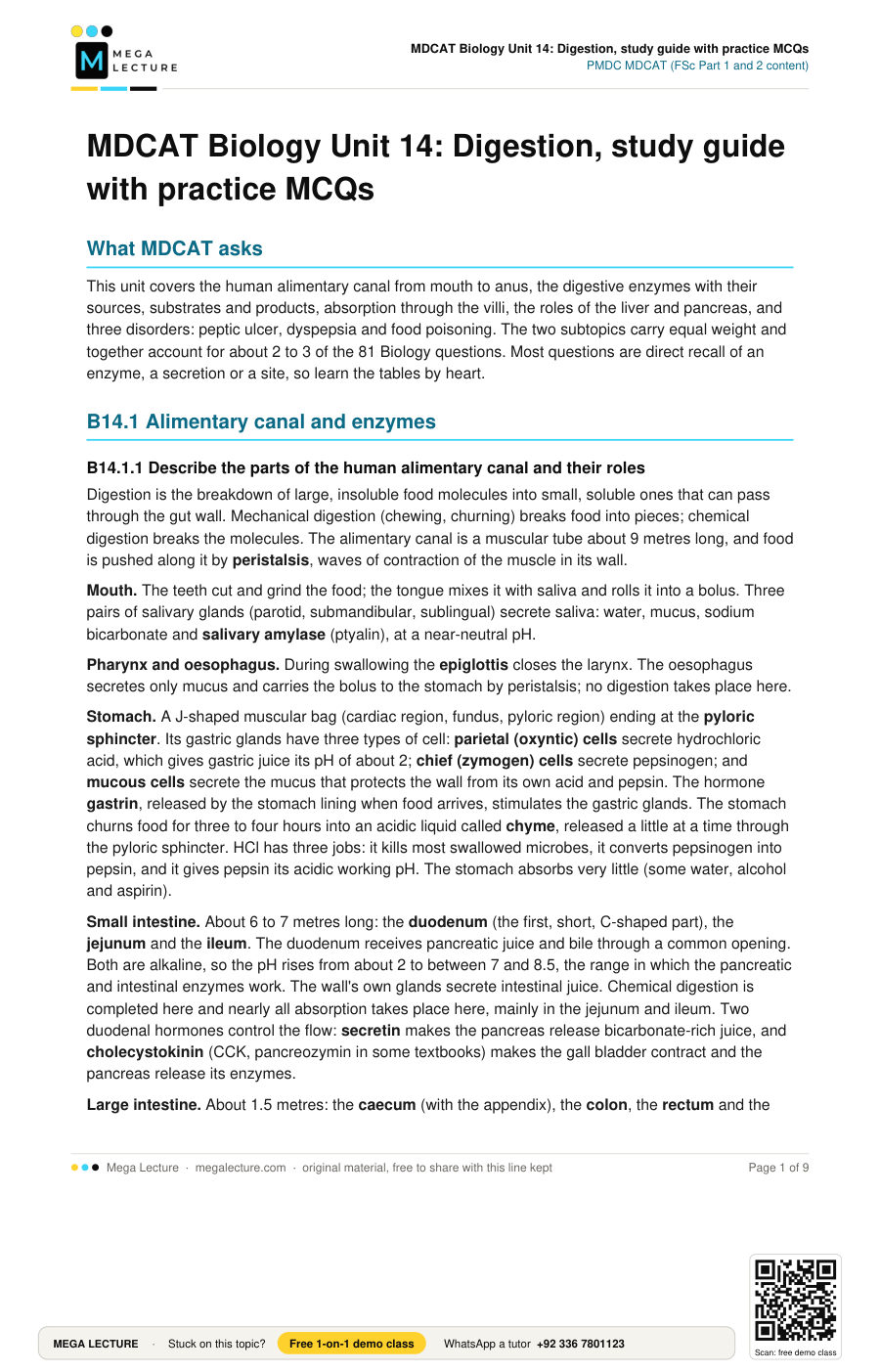
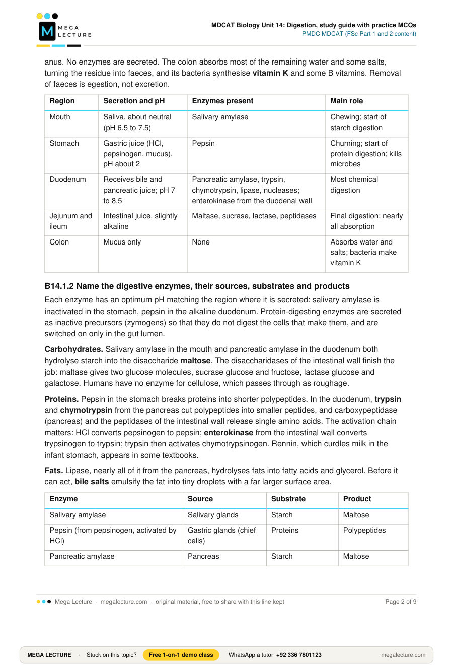
Opens inside this page. Prefer your own PDF app? Open the PDF.
Check yourself
5 quick questions on MDCAT Biology
Tap an answer to see if you are right, and why. Free, no sign-up.
Start the free testWas this note useful?
Full text of MDCAT Biology Unit 14: Digestion, study guide with practice MCQs
What MDCAT asks
This unit covers the human alimentary canal from mouth to anus, the digestive enzymes with their sources, substrates and products, absorption through the villi, the roles of the liver and pancreas, and three disorders: peptic ulcer, dyspepsia and food poisoning. The two subtopics carry equal weight and together account for about 2 to 3 of the 81 Biology questions. Most questions are direct recall of an enzyme, a secretion or a site, so learn the tables by heart.
B14.1 Alimentary canal and enzymes
B14.1.1 Describe the parts of the human alimentary canal and their roles
Digestion is the breakdown of large, insoluble food molecules into small, soluble ones that can pass through the gut wall. Mechanical digestion (chewing, churning) breaks food into pieces; chemical digestion breaks the molecules. The alimentary canal is a muscular tube about 9 metres long, and food is pushed along it by peristalsis, waves of contraction of the muscle in its wall.
Mouth. The teeth cut and grind the food; the tongue mixes it with saliva and rolls it into a bolus. Three pairs of salivary glands (parotid, submandibular, sublingual) secrete saliva: water, mucus, sodium bicarbonate and salivary amylase (ptyalin), at a near-neutral pH.
Pharynx and oesophagus. During swallowing the epiglottis closes the larynx. The oesophagus secretes only mucus and carries the bolus to the stomach by peristalsis; no digestion takes place here.
Stomach. A J-shaped muscular bag (cardiac region, fundus, pyloric region) ending at the pyloric sphincter. Its gastric glands have three types of cell: parietal (oxyntic) cells secrete hydrochloric acid, which gives gastric juice its pH of about 2; chief (zymogen) cells secrete pepsinogen; and mucous cells secrete the mucus that protects the wall from its own acid and pepsin. The hormone gastrin, released by the stomach lining when food arrives, stimulates the gastric glands. The stomach churns food for three to four hours into an acidic liquid called chyme, released a little at a time through the pyloric sphincter. HCl has three jobs: it kills most swallowed microbes, it converts pepsinogen into pepsin, and it gives pepsin its acidic working pH. The stomach absorbs very little (some water, alcohol and aspirin).
Small intestine. About 6 to 7 metres long: the duodenum (the first, short, C-shaped part), the jejunum and the ileum. The duodenum receives pancreatic juice and bile through a common opening. Both are alkaline, so the pH rises from about 2 to between 7 and 8.5, the range in which the pancreatic and intestinal enzymes work. The wall's own glands secrete intestinal juice. Chemical digestion is completed here and nearly all absorption takes place here, mainly in the jejunum and ileum. Two duodenal hormones control the flow: secretin makes the pancreas release bicarbonate-rich juice, and cholecystokinin (CCK, pancreozymin in some textbooks) makes the gall bladder contract and the pancreas release its enzymes.
Large intestine. About 1.5 metres: the caecum (with the appendix), the colon, the rectum and the anus. No enzymes are secreted. The colon absorbs most of the remaining water and some salts, turning the residue into faeces, and its bacteria synthesise vitamin K and some B vitamins. Removal of faeces is egestion, not excretion.
| Region | Secretion and pH | Enzymes present | Main role |
|---|---|---|---|
| Mouth | Saliva, about neutral (pH 6.5 to 7.5) | Salivary amylase | Chewing; start of starch digestion |
| Stomach | Gastric juice (HCl, pepsinogen, mucus), pH about 2 | Pepsin | Churning; start of protein digestion; kills microbes |
| Duodenum | Receives bile and pancreatic juice; pH 7 to 8.5 | Pancreatic amylase, trypsin, chymotrypsin, lipase, nucleases; enterokinase from the duodenal wall | Most chemical digestion |
| Jejunum and ileum | Intestinal juice, slightly alkaline | Maltase, sucrase, lactase, peptidases | Final digestion; nearly all absorption |
| Colon | Mucus only | None | Absorbs water and salts; bacteria make vitamin K |
B14.1.2 Name the digestive enzymes, their sources, substrates and products
Each enzyme has an optimum pH matching the region where it is secreted: salivary amylase is inactivated in the stomach, pepsin in the alkaline duodenum. Protein-digesting enzymes are secreted as inactive precursors (zymogens) so that they do not digest the cells that make them, and are switched on only in the gut lumen.
Carbohydrates. Salivary amylase in the mouth and pancreatic amylase in the duodenum both hydrolyse starch into the disaccharide maltose. The disaccharidases of the intestinal wall finish the job: maltase gives two glucose molecules, sucrase glucose and fructose, lactase glucose and galactose. Humans have no enzyme for cellulose, which passes through as roughage.
Proteins. Pepsin in the stomach breaks proteins into shorter polypeptides. In the duodenum, trypsin and chymotrypsin from the pancreas cut polypeptides into smaller peptides, and carboxypeptidase (pancreas) and the peptidases of the intestinal wall release single amino acids. The activation chain matters: HCl converts pepsinogen to pepsin; enterokinase from the intestinal wall converts trypsinogen to trypsin; trypsin then activates chymotrypsinogen. Rennin, which curdles milk in the infant stomach, appears in some textbooks.
Fats. Lipase, nearly all of it from the pancreas, hydrolyses fats into fatty acids and glycerol. Before it can act, bile salts emulsify the fat into tiny droplets with a far larger surface area.
| Enzyme | Source | Substrate | Product |
|---|---|---|---|
| Salivary amylase | Salivary glands | Starch | Maltose |
| Pepsin (from pepsinogen, activated by HCl) | Gastric glands (chief cells) | Proteins | Polypeptides |
| Pancreatic amylase | Pancreas | Starch | Maltose |
| Trypsin (from trypsinogen, activated by enterokinase) | Pancreas | Proteins, polypeptides | Smaller peptides |
| Chymotrypsin (activated by trypsin) | Pancreas | Polypeptides | Smaller peptides |
| Lipase | Pancreas (mainly) | Emulsified fats | Fatty acids + glycerol |
| Maltase | Intestinal wall | Maltose | Glucose + glucose |
| Sucrase | Intestinal wall | Sucrose | Glucose + fructose |
| Lactase | Intestinal wall | Lactose | Glucose + galactose |
| Peptidases | Intestinal wall (and pancreas) | Peptides | Amino acids |
Bile is not in the table because it contains no enzyme. It is made by the liver, stored and concentrated in the gall bladder, and released into the duodenum through the bile duct when fat arrives. It contains bile salts (emulsify fat), bile pigments (bilirubin and biliverdin, from the breakdown of haemoglobin; they colour the faeces), cholesterol and sodium bicarbonate (neutralises chyme).
MDCAT trap: bile and the "digestion of fat". Bile is a secretion, not an enzyme, and never breaks a chemical bond in a fat molecule. Emulsification is a physical change: one big globule becomes thousands of tiny droplets, so the surface exposed to lipase is far larger. The chemical digestion is done by lipase alone. Three rules:
- If the question asks "which enzyme digests fat", the answer is lipase, never bile.
- If it asks "what does bile do", the answers are emulsification, neutralising acid and giving an alkaline medium; "hydrolysis" is wrong.
- Bile is made in the liver and stored in the gall bladder; a person without a gall bladder still makes bile but cannot store a large dose for a fatty meal.
Likewise pepsinogen and trypsinogen digest nothing until activated: pepsinogen by HCl, trypsinogen by enterokinase. An option saying "trypsin is activated by HCl" has swapped them.
Worked example. A disease destroys the parietal cells of the gastric glands, so the stomach secretes no hydrochloric acid. Which step of protein digestion is lost, and why is protein digestion still nearly complete?
- Without HCl the stomach stays near neutral, so pepsinogen is not converted into pepsin, which could not work at that pH anyway. Gastric protein digestion stops.
- Trypsin is activated by enterokinase, not HCl, and then activates chymotrypsin; both work at the alkaline pH of the duodenum, and intestinal peptidases then release amino acids.
- Answer: only the pepsin step is lost; the pancreatic and intestinal proteases complete the job. The stomach also loses its acid defence against microbes.
B14.2 Absorption, liver and disorders
B14.2.1 Describe absorption in the small intestine and the role of villi
Absorption is the passage of the small, soluble products of digestion through the gut epithelium into the blood or lymph. Almost all of it happens in the small intestine; the stomach absorbs very little and the large intestine mainly water and salts. The small intestine is adapted for absorption in four ways:
- It is very long (6 to 7 metres), so food is in contact with the wall for hours.
- Its inner lining is thrown into circular folds.
- The folds carry millions of finger-like villi, each about 1 mm long.
- Each epithelial cell carries hundreds of microvilli (brush border), where the disaccharidases and peptidases sit. Together these give an internal surface about the size of a tennis court.
A villus has a wall one cell thick, a dense network of blood capillaries and a central lymph vessel, the lacteal. Its epithelial cells have many mitochondria, because much absorption is by active transport and needs ATP.
Two routes out of the villus.
- Glucose, amino acids, water-soluble vitamins, minerals and water enter the blood capillaries. Glucose and amino acids move by diffusion while their concentration in the gut is high, and by active transport once it falls below that in the blood. The capillaries drain into the hepatic portal vein, which carries absorbed food straight to the liver.
- Fatty acids and glycerol diffuse into the epithelial cells, helped by bile salts, and are rebuilt there into fats. Coated with protein as tiny chylomicrons, they pass into the lacteal; the lymph carries them into the blood near the heart, bypassing the liver at first. The fat-soluble vitamins A, D, E and K travel with the fats, so a person with poor bile flow becomes short of them.
| Absorbed substance | How it crosses the epithelium | Enters | Carried to |
|---|---|---|---|
| Glucose and other monosaccharides | Active transport and diffusion | Blood capillary | Liver by the hepatic portal vein |
| Amino acids | Active transport and diffusion | Blood capillary | Liver by the hepatic portal vein |
| Fatty acids, glycerol (rebuilt into fat) | Diffusion, then chylomicrons | Lacteal | Lymph, then blood near the heart |
| Vitamins A, D, E, K | Dissolved in fat droplets, with bile salts | Lacteal | Lymph |
| Water-soluble vitamins, salts, water | Diffusion, active transport, osmosis | Blood capillary | Liver by the hepatic portal vein |
B14.2.2 Describe the roles of the liver and pancreas, and digestive disorders (ulcer, dyspepsia, food poisoning)
The liver is the largest gland of the body and the first organ to receive absorbed food, because the hepatic portal vein brings blood from the gut directly to it. Its roles:
- Bile production: made continuously, stored in the gall bladder.
- Carbohydrate regulation: under insulin, liver cells store surplus glucose as glycogen; under glucagon they break glycogen down and release glucose, keeping blood glucose near 80 to 100 mg per 100 cm3.
- Protein metabolism: amino acids cannot be stored. Surplus ones are deaminated: the amino group is removed as toxic ammonia, which is at once converted into urea (ornithine cycle); the rest is used for energy or made into glucose or fat. The liver also makes the plasma proteins albumin, fibrinogen and prothrombin.
- Fat metabolism and detoxification: converts surplus carbohydrate to fat, makes cholesterol, and breaks down alcohol, drugs and other poisons.
- Storage and heat: stores glycogen, iron (from old red cells) and vitamins A, D and B12; its high metabolic rate produces much body heat.
The pancreas lies behind the stomach and is both an exocrine and an endocrine gland:
- The exocrine part (the acini) secretes pancreatic juice into the pancreatic duct, which opens into the duodenum. The juice is alkaline (sodium bicarbonate) and contains trypsinogen, chymotrypsinogen, procarboxypeptidase, amylase, lipase and nucleases; secretin and cholecystokinin stimulate its release.
- The endocrine part, the islets of Langerhans, secretes hormones directly into the blood: insulin from the beta cells (lowers blood glucose; glycogen is stored) and glucagon from the alpha cells (raises blood glucose; glycogen is broken down).
Peptic ulcer. An open sore in the lining of the stomach (gastric ulcer) or duodenum (duodenal ulcer), produced when acid and pepsin erode a wall whose mucus barrier has broken down. The commonest cause is the bacterium Helicobacter pylori, which lives in the mucus layer and damages it; aspirin-type painkillers, smoking, alcohol and stress also weaken the barrier or raise acid secretion. Symptoms are a burning upper-abdominal pain, often worse on an empty stomach, and in severe cases bleeding or perforation. Treatment: antibiotics against H. pylori plus acid-reducing drugs and antacids.
Dyspepsia is indigestion: discomfort in the upper abdomen after eating, with fullness, bloating, belching, heartburn and nausea. It is a set of symptoms rather than one disease, triggered by overeating, fatty or spicy food, stress, smoking and alcohol; persistent dyspepsia may be the first sign of an ulcer. It is managed by smaller meals, avoiding the triggers and antacids.
Food poisoning is illness caused by food or water contaminated with pathogenic microbes or their toxins. Salmonella (undercooked poultry and eggs), Campylobacter, Escherichia coli and Staphylococcus aureus cause most cases; Clostridium botulinum, which grows in badly canned food, makes the toxin of botulism, a rare form that paralyses muscles. Symptoms appear within hours to two days: nausea, vomiting, cramps, diarrhoea, sometimes fever. The main danger is dehydration, so treatment is fluid and salt replacement; prevention is thorough cooking, refrigeration and clean hands. The FSc textbook also lists obesity, anorexia nervosa, bulimia nervosa and piles; know their one-line definitions.
| Disorder | What it is | Usual cause | Key symptom |
|---|---|---|---|
| Peptic ulcer | Open sore in the wall of the stomach or duodenum | Helicobacter pylori; aspirin-type drugs; smoking, stress | Burning upper-abdominal pain, often on an empty stomach |
| Dyspepsia | Indigestion: a group of symptoms after eating | Overeating, fatty food, stress; sometimes an ulcer | Fullness, bloating, heartburn, nausea |
| Food poisoning | Illness from microbes or toxins in food | Salmonella, Campylobacter, E. coli, C. botulinum toxin | Vomiting, diarrhoea, cramps; risk of dehydration |
MDCAT trap: who secretes what. Examiners attach the right product to the wrong organ. The liver makes bile and stores glycogen; it secretes no digestive enzyme and no hormone. Insulin and glucagon come from the islets of Langerhans in the pancreas; the liver only obeys them, storing glucose as glycogen under insulin and releasing it under glucagon. Four checks:
- "Enzyme" in the stem: the source is the salivary glands, stomach, pancreas or intestinal wall, never the liver.
- "Hormone" in the stem: gastrin from the stomach wall, secretin and cholecystokinin from the duodenal wall, insulin and glucagon from the pancreatic islets; none from the liver.
- "Stores" in the stem: the liver stores glycogen, iron and vitamins; the gall bladder stores bile; the pancreas stores nothing.
- "Into a duct" means exocrine (pancreatic juice, bile); "into the blood" means endocrine (insulin, glucagon). The pancreas does both.
Worked example. A blockage of the bile duct stops bile reaching the duodenum, although the pancreas is healthy. Predict the effect on (a) the faeces, (b) absorption of vitamin K and (c) blood clotting.
- (a) Bile pigments normally colour the faeces brown, so they become pale. They are also greasy, because without emulsification lipase works slowly and much fat passes out undigested.
- (b) Vitamin K is fat-soluble and is absorbed only in emulsified fat droplets, which need bile salts, so its absorption falls sharply even though the ileum is normal.
- (c) The liver needs vitamin K to make prothrombin, so clotting slows and the person bleeds easily.
- Answer: pale, fatty faeces; poor vitamin K absorption; prolonged bleeding, through the chain bile → fat absorption → vitamin K → prothrombin.
Practice MCQs
- Which digestive enzyme is secreted in an inactive form and activated by HCl?
A. Trypsin
B. Lipase
C. Pepsin
D. Salivary amylase - A patient has had the gall bladder surgically removed. After a very fatty meal, the digestion of which food class is most likely to be slower than normal?
A. Fats
B. Starch
C. Proteins
D. Sucrose - Salivary amylase stops acting soon after the food bolus enters the stomach. The best explanation is that
A. starch has already been fully digested in the mouth
B. the temperature of the stomach is too low for the enzyme
C. the enzyme requires bile salts, which are absent in the stomach
D. the acidic pH of the stomach is far from the enzyme's optimum - Which row correctly matches an enzyme with its source and product?
A. Pepsin: pancreas: amino acids
B. Lactase: intestinal wall: glucose and galactose
C. Trypsin: stomach: peptides
D. Lipase: liver: fatty acids and glycerol - Trypsinogen released into the duodenum is converted into active trypsin by
A. enterokinase from the intestinal wall
B. hydrochloric acid from the stomach
C. bile salts from the gall bladder
D. secretin from the duodenal wall - A drug blocks ATP production in the epithelial cells of the villi. The absorption of which substance would be reduced most?
A. Fatty acids
B. Glycerol
C. Glucose
D. Vitamin A - Which absorbed product leaves the villus in the lacteal rather than in a blood capillary?
A. Glucose
B. Amino acids
C. Vitamin C
D. Fats rebuilt from fatty acids and glycerol - A person eats a very large meal of boiled eggs with almost no carbohydrate. A few hours later the urea concentration of the blood leaving the liver has risen. Which liver process explains this?
A. Breakdown of glycogen into glucose
B. Deamination of surplus amino acids
C. Emulsification of fats by bile
D. Conversion of glucose into glycogen - Which statement about the pancreas is correct?
A. It secretes bile into the duodenum through the pancreatic duct
B. Its islets of Langerhans release insulin directly into the blood, not into a duct
C. It stores surplus glucose as glycogen after a meal
D. It secretes pepsinogen, which is activated in the duodenum - A peptic ulcer is most directly the result of
A. acid and pepsin eroding a region of the stomach or duodenal wall that has lost its mucus protection
B. toxins produced by bacteria growing in badly stored food
C. the absence of bile salts in the duodenum
D. blockage of the pancreatic duct by a tumour
1. C. Chief cells secrete pepsinogen and HCl converts it into pepsin. Trypsin is activated by enterokinase; lipase and amylase are secreted active.
2. A. Without the gall bladder only a trickle of bile reaches the duodenum, so fat is poorly emulsified and lipase works slowly. The other three do not need bile.
3. D. Salivary amylase works near pH 7; at pH 2 it is denatured. Starch is not fully digested in the mouth, and temperature is the same in both.
4. B. Lactase from the intestinal wall splits lactose into glucose and galactose. Pepsin comes from the stomach, and trypsin and lipase from the pancreas, not the liver.
5. A. Enterokinase from the intestinal wall converts trypsinogen into trypsin, which then activates the other pancreatic zymogens; HCl activates pepsinogen.
6. C. Glucose is absorbed largely by active transport, which needs ATP; fatty acids, glycerol and vitamin A enter by diffusion.
7. D. Fatty acids and glycerol are rebuilt into fats in the epithelial cells and enter the lacteal; glucose, amino acids and vitamin C enter the blood capillaries.
8. B. Egg protein gives amino acids, which the liver cannot store; the surplus is deaminated and the amino groups become urea. No other option makes urea.
9. B. The islets are the endocrine part of the pancreas and release insulin and glucagon into the blood. Bile and glycogen belong to the liver, pepsinogen to the stomach.
10. A. An ulcer forms when the mucus barrier fails (usually from Helicobacter pylori or aspirin-type drugs) and acid and pepsin attack the wall. B describes food poisoning; C and D affect fat digestion, not the wall.
Before the test: 12 facts to recite
- Order: mouth → oesophagus → stomach → duodenum → jejunum → ileum → caecum → colon → rectum → anus; food moves by peristalsis.
- Saliva is about neutral, gastric juice about pH 2, the duodenum alkaline because of bile and pancreatic bicarbonate.
- Gastric glands: parietal cells make HCl, chief cells make pepsinogen, mucous cells make protective mucus; gastrin stimulates them.
- HCl kills microbes, converts pepsinogen into pepsin and gives pepsin its working pH.
- Amylase: starch → maltose. Maltase: maltose → glucose + glucose. Sucrase: sucrose → glucose + fructose. Lactase: lactose → glucose + galactose.
- Pepsin (stomach), trypsin and chymotrypsin (pancreas) cut proteins into peptides; intestinal peptidases release amino acids; enterokinase activates trypsinogen.
- Lipase, almost all from the pancreas, hydrolyses fats to fatty acids and glycerol; bile only emulsifies fat and contains no enzyme.
- Bile is made in the liver and stored in the gall bladder; it contains bile salts, pigments, cholesterol and bicarbonate.
- Villi and microvilli give a huge surface; a villus has a thin wall, blood capillaries and a central lacteal.
- Glucose and amino acids go to the blood capillaries and the hepatic portal vein; rebuilt fats and vitamins A, D, E and K go to the lacteal and the lymph.
- The liver stores glycogen, deaminates amino acids to make urea, detoxifies poisons and makes plasma proteins; it secretes no enzyme and no hormone.
- The pancreas is exocrine (pancreatic juice into a duct) and endocrine (insulin and glucagon into the blood). Peptic ulcer: acid and pepsin on an unprotected wall, usually with Helicobacter pylori; dyspepsia: indigestion; food poisoning: microbes or toxins in food.
