
MDCAT · Biology (PMDC curriculum) · Unit 5: Coordination and control
MDCAT Biology Unit 5: Coordination and control, study guide with practice MCQs
Need more than one subject? All-notes pass: unlimited downloads of every note for a year: unlimited downloads of every note on the site, PKR 999 a year. All packs and courses.
Opens inside this page. Prefer your own PDF app? Open the PDF.
Check yourself
5 quick questions on MDCAT Biology
Tap an answer to see if you are right, and why. Free, no sign-up.
Start the free testWas this note useful?
Full text of MDCAT Biology Unit 5: Coordination and control, study guide with practice MCQs
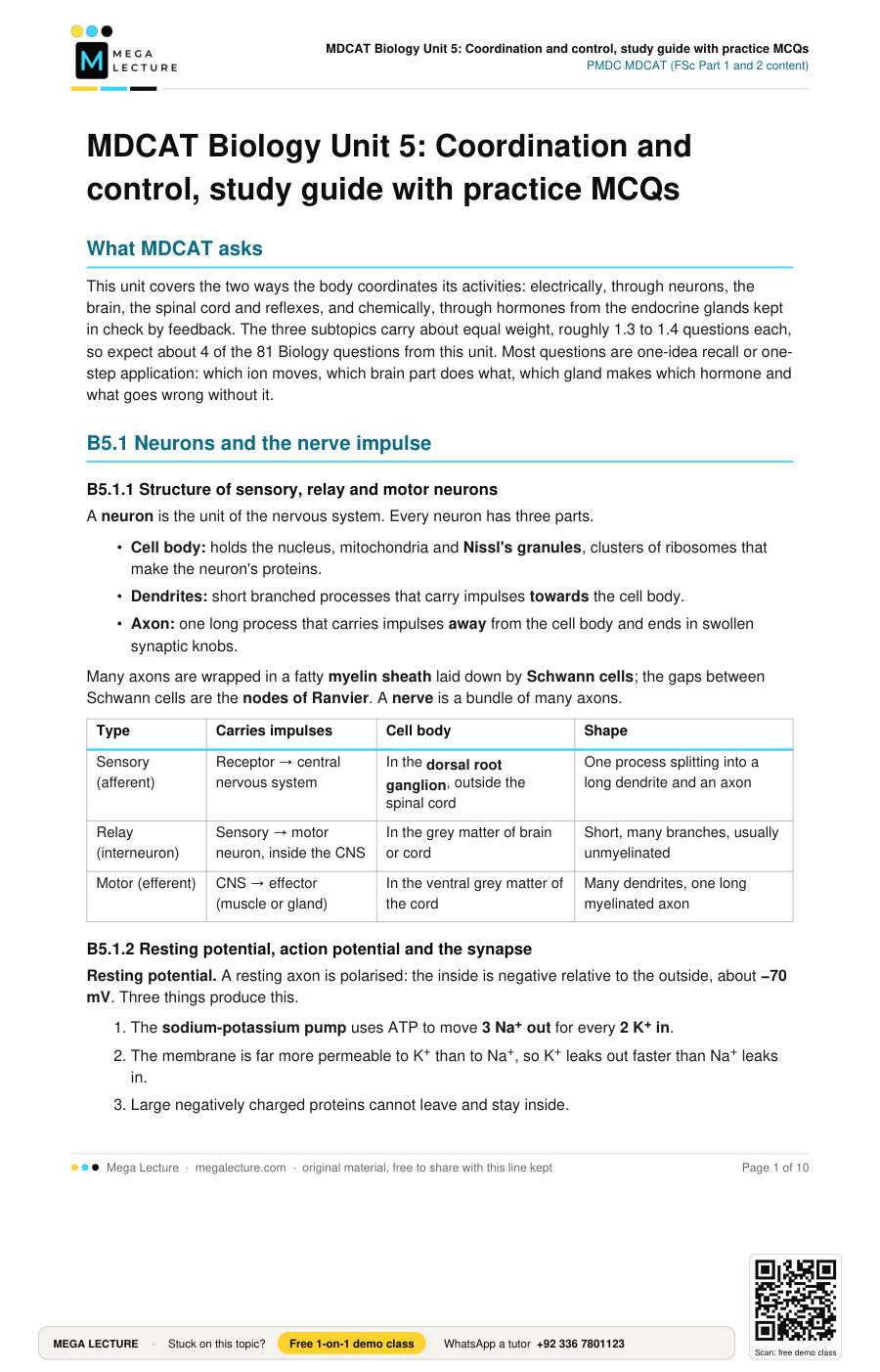
What MDCAT asks
This unit covers the two ways the body coordinates its activities: electrically, through neurons, the brain, the spinal cord and reflexes, and chemically, through hormones from the endocrine glands kept in check by feedback. The three subtopics carry about equal weight, roughly 1.3 to 1.4 questions each, so expect about 4 of the 81 Biology questions from this unit. Most questions are one-idea recall or one-step application: which ion moves, which brain part does what, which gland makes which hormone and what goes wrong without it.
B5.1 Neurons and the nerve impulse
B5.1.1 Structure of sensory, relay and motor neurons
A neuron is the unit of the nervous system. Every neuron has three parts.
- Cell body: holds the nucleus, mitochondria and Nissl's granules, clusters of ribosomes that make the neuron's proteins.
- Dendrites: short branched processes that carry impulses towards the cell body.
- Axon: one long process that carries impulses away from the cell body and ends in swollen synaptic knobs.
Many axons are wrapped in a fatty myelin sheath laid down by Schwann cells; the gaps between Schwann cells are the nodes of Ranvier. A nerve is a bundle of many axons.
| Type | Carries impulses | Cell body | Shape |
|---|---|---|---|
| Sensory (afferent) | Receptor → central nervous system | In the dorsal root ganglion, outside the spinal cord | One process splitting into a long dendrite and an axon |
| Relay (interneuron) | Sensory → motor neuron, inside the CNS | In the grey matter of brain or cord | Short, many branches, usually unmyelinated |
| Motor (efferent) | CNS → effector (muscle or gland) | In the ventral grey matter of the cord | Many dendrites, one long myelinated axon |
B5.1.2 Resting potential, action potential and the synapse
Resting potential. A resting axon is polarised: the inside is negative relative to the outside, about −70 mV. Three things produce this.
- The sodium-potassium pump uses ATP to move 3 Na+ out for every 2 K+ in.
- The membrane is far more permeable to K+ than to Na+, so K+ leaks out faster than Na+ leaks in.
- Large negatively charged proteins cannot leave and stay inside.
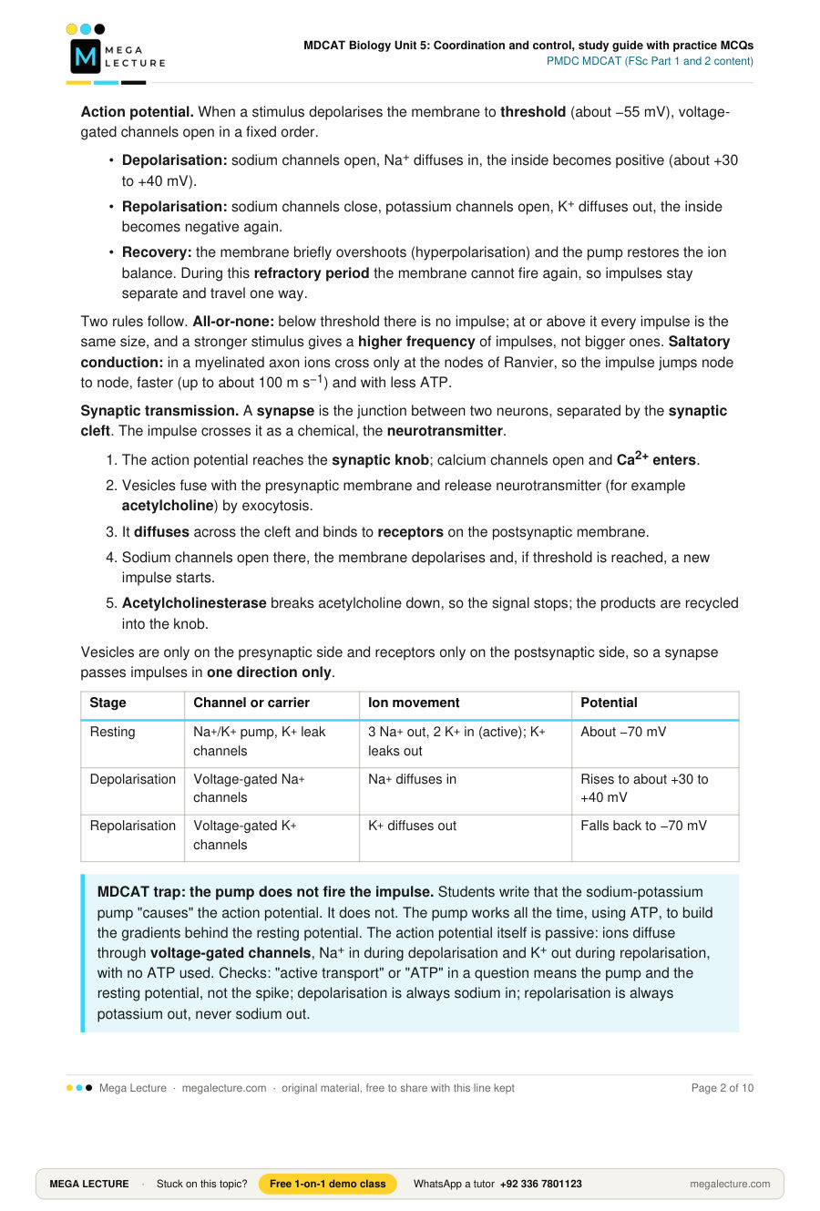
Action potential. When a stimulus depolarises the membrane to threshold (about −55 mV), voltage-gated channels open in a fixed order.
- Depolarisation: sodium channels open, Na+ diffuses in, the inside becomes positive (about +30 to +40 mV).
- Repolarisation: sodium channels close, potassium channels open, K+ diffuses out, the inside becomes negative again.
- Recovery: the membrane briefly overshoots (hyperpolarisation) and the pump restores the ion balance. During this refractory period the membrane cannot fire again, so impulses stay separate and travel one way.
Two rules follow. All-or-none: below threshold there is no impulse; at or above it every impulse is the same size, and a stronger stimulus gives a higher frequency of impulses, not bigger ones. Saltatory conduction: in a myelinated axon ions cross only at the nodes of Ranvier, so the impulse jumps node to node, faster (up to about 100 m s−1) and with less ATP.
Synaptic transmission. A synapse is the junction between two neurons, separated by the synaptic cleft. The impulse crosses it as a chemical, the neurotransmitter.
- The action potential reaches the synaptic knob; calcium channels open and Ca2+ enters.
- Vesicles fuse with the presynaptic membrane and release neurotransmitter (for example acetylcholine) by exocytosis.
- It diffuses across the cleft and binds to receptors on the postsynaptic membrane.
- Sodium channels open there, the membrane depolarises and, if threshold is reached, a new impulse starts.
- Acetylcholinesterase breaks acetylcholine down, so the signal stops; the products are recycled into the knob.
Vesicles are only on the presynaptic side and receptors only on the postsynaptic side, so a synapse passes impulses in one direction only.
| Stage | Channel or carrier | Ion movement | Potential |
|---|---|---|---|
| Resting | Na+/K+ pump, K+ leak channels | 3 Na+ out, 2 K+ in (active); K+ leaks out | About −70 mV |
| Depolarisation | Voltage-gated Na+ channels | Na+ diffuses in | Rises to about +30 to +40 mV |
| Repolarisation | Voltage-gated K+ channels | K+ diffuses out | Falls back to −70 mV |
MDCAT trap: the pump does not fire the impulse. Students write that the sodium-potassium pump "causes" the action potential. It does not. The pump works all the time, using ATP, to build the gradients behind the resting potential. The action potential itself is passive: ions diffuse through voltage-gated channels, Na+ in during depolarisation and K+ out during repolarisation, with no ATP used. Checks: "active transport" or "ATP" in a question means the pump and the resting potential, not the spike; depolarisation is always sodium in; repolarisation is always potassium out, never sodium out.
Worked example. A drug blocks the acetylcholine receptors on the muscle membrane at a neuromuscular junction but leaves the motor neuron untouched. The nerve is stimulated above threshold. What happens?
- The action potential travels normally along the axon.
- Ca2+ enters the knob and vesicles release acetylcholine as usual.
- Acetylcholine diffuses across but cannot bind, because the receptors are blocked.
- The muscle membrane does not depolarise, so there is no contraction.
Answer: the impulse reaches the synapse but transmission fails at the postsynaptic receptors, so the muscle is paralysed.
B5.2 The nervous system
B5.2.1 Parts of the human brain and spinal cord and their functions
The central nervous system (CNS) is the brain and spinal cord. The peripheral nervous system (PNS) is the 12 pairs of cranial nerves and 31 pairs of spinal nerves. The PNS has a somatic division (voluntary control of skeletal muscle) and an autonomic division (involuntary control of smooth muscle, heart and glands), split into sympathetic and parasympathetic systems. The CNS is protected by bone, the meninges and cerebrospinal fluid.
| Region | Part | Main functions |
|---|---|---|
| Forebrain | Cerebrum (two hemispheres; outer cortex of grey matter) | Intelligence, memory, speech, voluntary movement, interpretation of sensations. Each hemisphere controls the opposite side of the body; the corpus callosum joins the two |
| Forebrain | Thalamus | Relay station that sorts sensory impulses and sends them to the correct area of the cortex |
| Forebrain | Hypothalamus and limbic system (amygdala, hippocampus) | Hypothalamus: temperature, hunger, thirst, water balance, control of the pituitary. Amygdala: emotion. Hippocampus: new memories |
| Midbrain | Midbrain | Relays impulses between forebrain and hindbrain; reflex centres for sight and hearing |
| Hindbrain | Cerebellum | Balance, posture, muscle tone, fine coordination of voluntary movement; it smooths movements, it does not start them |
| Hindbrain | Pons | Bridge of fibres linking cerebellum, cerebrum and medulla |
| Hindbrain | Medulla oblongata | Vital reflex centres: breathing, heartbeat, blood pressure, swallowing, vomiting |
The spinal cord in cross section has an H-shaped core of grey matter (cell bodies and synapses) surrounded by white matter (myelinated axons running to and from the brain). Note the reversal: in the cerebrum the grey matter is outside; in the cord it is inside. Each spinal nerve joins the cord by two roots: the dorsal root carries sensory fibres in and bears the dorsal root ganglion; the ventral root carries motor fibres out.
B5.2.2 The reflex arc, receptors and common nervous disorders
A reflex is a rapid, automatic, involuntary response to a stimulus that does not need a decision by the cerebrum. Reflexes protect the body (withdrawal, blinking, coughing) and keep posture (knee jerk). The pathway is the reflex arc:
- Receptor detects the stimulus and generates an impulse.
- Sensory neuron carries it into the spinal cord through the dorsal root.
- Relay neuron in the grey matter links sensory and motor neurons and sends a branch up to the brain. (The knee jerk has no relay neuron.)
- Motor neuron leaves through the ventral root.
- Effector, a muscle or gland, responds.
The two autonomic systems act on the same organs with opposite effects. Sympathetic (fight or flight): faster heart, higher blood pressure, dilated pupils and bronchioles, slower digestion, raised blood glucose. Parasympathetic (rest and digest): slower heart, constricted pupils, active digestion.
Receptors convert the energy of a stimulus into nerve impulses and are named by the stimulus they detect.
- Chemoreceptors: chemicals; taste buds, olfactory cells, CO2 sensors in the arteries.
- Mechanoreceptors: touch, pressure, stretch, sound; skin corpuscles, hair cells of the cochlea.
- Photoreceptors: light; rods and cones of the retina.
- Thermoreceptors: heat and cold; skin and hypothalamus.
- Nociceptors: pain; free nerve endings.
Common nervous disorders.
- Parkinson's disease: death of dopamine-producing neurons in the midbrain, so dopamine is lacking. Resting tremor, rigidity, slow movement. Treated with L-dopa, a precursor of dopamine.
- Epilepsy: sudden bursts of uncontrolled electrical activity in the cerebral cortex cause seizures (fits), controlled with anticonvulsant drugs.
- Alzheimer's disease: progressive degeneration of cortical neurons with protein plaques and a fall in acetylcholine: loss of recent memory, then confusion; the commonest dementia of old age.
MDCAT trap: look-alike names. Two pairs are swapped more than any others. First, cerebrum and cerebellum. The cerebrum is the large folded thinking brain: intelligence, memory, voluntary movement, the senses. The cerebellum is the smaller structure beneath it: balance, posture and coordination of the movements the cerebrum has ordered. Staggering means cerebellar damage; loss of speech or memory means cerebral damage. Second, dorsal and ventral roots. Dorsal is sensory, in, with the ganglion; ventral is motor, out, no ganglion. Cutting the dorsal root removes sensation but the muscle can still be moved; cutting the ventral root paralyses the muscle but sensation remains.
Worked example. A student steps on a pin and lifts the foot before feeling any pain. Explain the sequence and why the pain comes afterwards.
- Nociceptors in the skin generate impulses.
- A sensory neuron carries them into the cord through the dorsal root; its cell body is in the dorsal root ganglion.
- In the grey matter it synapses with a relay neuron, which synapses with a motor neuron.
- The motor neuron leaves through the ventral root and the leg flexors contract. Only two synapses are crossed, so this is very fast.
- The relay neuron also sends impulses up the white matter to the thalamus and cerebral cortex, where pain is interpreted.
Answer: the withdrawal is a spinal reflex that does not wait for the brain; pain is felt in the cerebrum, which the impulses reach later by a longer path.
B5.3 Chemical coordination
B5.3.1 Endocrine glands, their hormones and effects
Hormones are organic molecules (proteins, amines or steroids) secreted in tiny amounts by endocrine glands. These glands are ductless: the hormone passes straight into the blood, which carries it everywhere, but only target cells with the matching receptor respond. Compared with nervous control, hormonal control is slower, lasts longer and acts on many organs at once instead of one precise effector.
The pituitary is the master gland because several of its hormones control other glands, but it is itself controlled by the hypothalamus, which links the nervous and endocrine systems.
| Gland | Hormone | Main effect |
|---|---|---|
| Anterior pituitary | Growth hormone | Growth of bones and soft tissues, protein synthesis |
| Anterior pituitary | TSH | Stimulates thyroid to release thyroxine |
| Anterior pituitary | ACTH | Stimulates adrenal cortex to release cortisol |
| Anterior pituitary | FSH and LH | Follicle growth and ovulation; sperm production and testosterone secretion |
| Anterior pituitary | Prolactin | Milk production |
| Posterior pituitary (stores hormones made in the hypothalamus) | ADH (vasopressin) | Water reabsorption in kidney collecting ducts; concentrated urine |
| Posterior pituitary | Oxytocin | Uterine contraction in labour; milk ejection |
| Thyroid | Thyroxine (needs iodine) | Raises metabolic rate, heat production, growth and mental development |
| Thyroid | Calcitonin | Lowers blood Ca2+ by depositing it in bone |
| Parathyroid | Parathormone | Raises blood Ca2+ from bone, gut and kidney |
| Pancreas, islets of Langerhans | Insulin (β cells) | Lowers blood glucose: liver and muscle make glucose → glycogen |
| Pancreas, islets of Langerhans | Glucagon (α cells) | Raises blood glucose: liver makes glycogen → glucose |
| Adrenal medulla | Adrenaline, noradrenaline | Emergency: faster heart, dilated pupils and bronchioles, glycogen → glucose |
| Adrenal cortex | Cortisol | Raises blood glucose during long-term stress; anti-inflammatory |
| Adrenal cortex | Aldosterone | Na+ reabsorption in the kidney, raising blood volume and pressure |
| Testes | Testosterone | Sperm production, male secondary characteristics |
| Ovaries | Oestrogen, progesterone | Female secondary characteristics, uterine lining; maintenance of pregnancy |
Note: ADH and oxytocin are made in the hypothalamus and only stored by the posterior pituitary; thyroxine is the only hormone that contains iodine.
B5.3.2 Feedback control of hormone secretion and disorders of hormone secretion
Hormone levels are held within narrow limits by negative feedback: a rise in the hormone, or in the condition it produces, switches off further secretion; a fall switches it on again. The system corrects its own deviations and keeps the body steady.
Thyroxine. Hypothalamus releases TRH → anterior pituitary releases TSH → thyroid releases thyroxine. High thyroxine inhibits the hypothalamus and the pituitary, so TSH falls and the thyroid slows; low thyroxine lifts the inhibition and TSH rises. Cortisol (through ACTH) is controlled the same way.
Blood glucose. After a meal glucose rises, β cells release insulin, glucose is stored as glycogen and the level falls. Between meals glucose falls, α cells release glucagon, the liver breaks glycogen to glucose and the level rises. Two hormones with opposite (antagonistic) effects give control in both directions. Calcitonin and parathormone control blood Ca2+ in the same way.
Positive feedback is rare and drives a process to completion, as oxytocin does in labour.
Disorders arise when a gland secretes too much (hypersecretion) or too little (hyposecretion).
| Hormone | Too little | Too much |
|---|---|---|
| Growth hormone | Childhood: dwarfism, small with normal proportions | Childhood: gigantism. Adult, after growth plates close: acromegaly, enlarged hands, feet and jaw |
| Thyroxine | Childhood: cretinism, stunted growth and mental retardation. Adult: myxoedema, low metabolic rate, weight gain, lethargy. Low dietary iodine: goitre, a swollen thyroid | Graves' disease: high metabolic rate, weight loss, rapid heartbeat, nervousness, bulging eyes |
| Insulin | Diabetes mellitus: high blood glucose, glucose in urine, large urine volume, thirst, weight loss. Type 1: β cells destroyed. Type 2: cells stop responding to insulin | Hypoglycaemia: weakness, sweating, loss of consciousness |
| ADH | Diabetes insipidus: very large volumes of dilute urine, constant thirst, no glucose in urine, normal blood glucose | Water retention |
| Adrenal cortex hormones | Addison's disease: low blood glucose and sodium, low blood pressure, weakness | Cushing's syndrome: high blood glucose, moon face, high blood pressure |
MDCAT trap: two diseases called diabetes. Diabetes only means passing a lot of urine. Both conditions do that and both cause thirst, but the hormone and the urine differ. Diabetes mellitus ("sweet") is a lack of insulin, or of the response to it: blood glucose is high, the kidney cannot reabsorb it all, so glucose appears in the urine and draws water with it. Diabetes insipidus ("tasteless") is a lack of ADH: blood glucose is normal, the urine has no glucose, but the collecting ducts reabsorb too little water, so the urine is huge in volume and very dilute. Test: glucose, pancreas or insulin in the question means mellitus; ADH, posterior pituitary or water reabsorption means insipidus. One more: insulin comes from β cells and glucagon from α cells, not the reverse.
Worked example. A 40-year-old woman has gained weight, feels cold and tired, has a slow pulse and puffy skin. Blood tests show low thyroxine and high TSH. Where is the fault, and why is TSH high?
- Low metabolic rate, cold intolerance, lethargy and weight gain in an adult are hypothyroidism: myxoedema.
- Under negative feedback, low thyroxine removes the inhibition on the pituitary, so TSH rises to drive the thyroid harder.
- TSH is high, so the pituitary is working; the thyroid receives its signal but fails to respond, so the fault is in the thyroid itself (or in lack of iodine).
Answer: myxoedema from a failing thyroid; high TSH is the pituitary's normal feedback response to low thyroxine. If the pituitary were at fault, TSH would be low too.
Practice MCQs
- Which part of a neuron carries impulses towards the cell body?
A. Axon
B. Dendrite
C. Synaptic knob
D. Node of Ranvier - During repolarisation of an axon membrane, which event takes place?
A. Na+ diffuses in through voltage-gated channels
B. The sodium-potassium pump moves Na+ into the axon
C. K+ diffuses into the axon
D. K+ diffuses out through voltage-gated channels - An insecticide inhibits acetylcholinesterase at synapses. What is the most likely immediate effect?
A. Acetylcholine remains in the cleft and keeps stimulating the postsynaptic membrane
B. No neurotransmitter is released from the synaptic knob
C. Impulses begin to pass backwards across the synapse
D. Ca2+ cannot enter the synaptic knob - An axon is stimulated with a stimulus twice as strong as threshold. Compared with a stimulus exactly at threshold:
A. each action potential is twice as large
B. each action potential travels twice as fast
C. more action potentials are produced per second, each of the same size
D. the resting potential becomes more negative - A patient can stand and move his limbs but staggers when walking and cannot touch his nose with his eyes closed. The region most likely damaged is the:
A. medulla oblongata
B. cerebellum
C. thalamus
D. hypothalamus - In a cross section of the spinal cord, the cell bodies of relay and motor neurons are found in the:
A. white matter
B. ventral root
C. meninges
D. central H-shaped grey matter - Which sequence correctly shows the path of impulses in a withdrawal reflex?
A. Receptor → sensory neuron → relay neuron → motor neuron → effector
B. Receptor → motor neuron → relay neuron → sensory neuron → effector
C. Receptor → sensory neuron → cerebrum → motor neuron → effector
D. Effector → sensory neuron → relay neuron → motor neuron → receptor - A tumour of the thyroid releases thyroxine continuously. Under negative feedback, the anterior pituitary would be expected to:
A. increase TSH secretion
B. increase ACTH secretion
C. decrease TSH secretion
D. decrease growth hormone secretion - Half an hour after a meal rich in carbohydrate, which response is correct?
A. α cells release glucagon and the liver converts glucose → glycogen
B. β cells release insulin and the liver converts glycogen → glucose
C. α cells release insulin and the liver converts glucose → glycogen
D. β cells release insulin and liver and muscle convert glucose → glycogen - A patient produces large volumes of urine, is constantly thirsty and is losing weight. Glucose is present in the urine. The hormone that is deficient is normally secreted by the:
A. β cells of the islets of Langerhans
B. posterior pituitary
C. adrenal cortex
D. α cells of the islets of Langerhans
1. B. Dendrites conduct impulses towards the cell body; the axon, with its knob and nodes, conducts away from it.
2. D. Repolarisation is K+ diffusing out through voltage-gated potassium channels. Sodium entry is depolarisation, and the pump moves Na+ out, not in.
3. A. Acetylcholinesterase normally destroys acetylcholine in the cleft; with it blocked, the transmitter persists and keeps stimulating the postsynaptic membrane.
4. C. All-or-none: above threshold every action potential has the same size and speed; a stronger stimulus is coded as a higher frequency.
5. B. Balance and coordination are cerebellar functions; movements can still be started, so the cerebrum is intact.
6. D. Grey matter, which contains cell bodies, forms the H-shaped core of the cord; the white matter around it is myelinated axons.
7. A. Receptor → sensory neuron (dorsal root) → relay neuron → motor neuron (ventral root) → effector; the cerebrum is not in the arc.
8. C. High thyroxine inhibits the hypothalamus and anterior pituitary, so TSH secretion falls.
9. D. Rising glucose stimulates β cells to release insulin, which drives glucose into cells and its storage as glycogen in liver and muscle.
10. A. Glucose in the urine with thirst and weight loss is diabetes mellitus, a lack of insulin from the β cells. Lack of ADH (posterior pituitary) would give dilute urine with no glucose.
Before the test: 12 facts to recite
- Dendrites carry impulses towards the cell body, the axon away from it; sensory cell bodies sit in the dorsal root ganglion.
- Resting potential is about −70 mV, inside negative: pump 3 Na+ out and 2 K+ in, high K+ permeability, trapped negative ions.
- Depolarisation is Na+ diffusing in; repolarisation is K+ diffusing out; the pump only restores the gradients afterwards.
- All-or-none: no impulse below threshold, same-sized impulse above it; stimulus strength is coded as frequency.
- Myelin from Schwann cells gives saltatory conduction, node to node, faster and cheaper in ATP.
- Synapse: Ca2+ in → vesicles release neurotransmitter → diffusion across the cleft → receptors → new impulse; acetylcholinesterase ends the signal; one way only.
- CNS is brain plus spinal cord; PNS is 12 pairs of cranial and 31 pairs of spinal nerves.
- Cerebrum: thinking, memory, voluntary movement, opposite side of the body. Cerebellum: balance and coordination. Medulla: breathing, heartbeat, blood pressure. Hypothalamus: temperature, hunger, thirst, pituitary.
- Spinal cord: grey matter inside (H shape), white matter outside; dorsal root is sensory in, ventral root is motor out.
- Reflex arc: receptor → sensory neuron → relay neuron → motor neuron → effector. Sympathetic prepares for emergency; parasympathetic restores rest.
- Anterior pituitary: GH, TSH, ACTH, FSH, LH, prolactin. Posterior pituitary stores ADH and oxytocin made in the hypothalamus. Thyroxine needs iodine; insulin (β) lowers glucose, glucagon (α) raises it.
- Growth hormone: dwarfism, gigantism, acromegaly. Thyroxine: cretinism, myxoedema, Graves' disease. No insulin: diabetes mellitus (glucose in urine). No ADH: diabetes insipidus (dilute urine, no glucose). Adrenal cortex low: Addison's; high: Cushing's.
