
MDCAT · Biology (PMDC curriculum) · Unit 3: Biological molecules
MDCAT Biology Unit 3: Biological molecules, study guide with practice MCQs
Need more than one subject? All-notes pass: unlimited downloads of every note for a year: unlimited downloads of every note on the site, PKR 999 a year. All packs and courses.
Opens inside this page. Prefer your own PDF app? Open the PDF.
Check yourself
5 quick questions on MDCAT Biology
Tap an answer to see if you are right, and why. Free, no sign-up.
Start the free testWas this note useful?
Full text of MDCAT Biology Unit 3: Biological molecules, study guide with practice MCQs
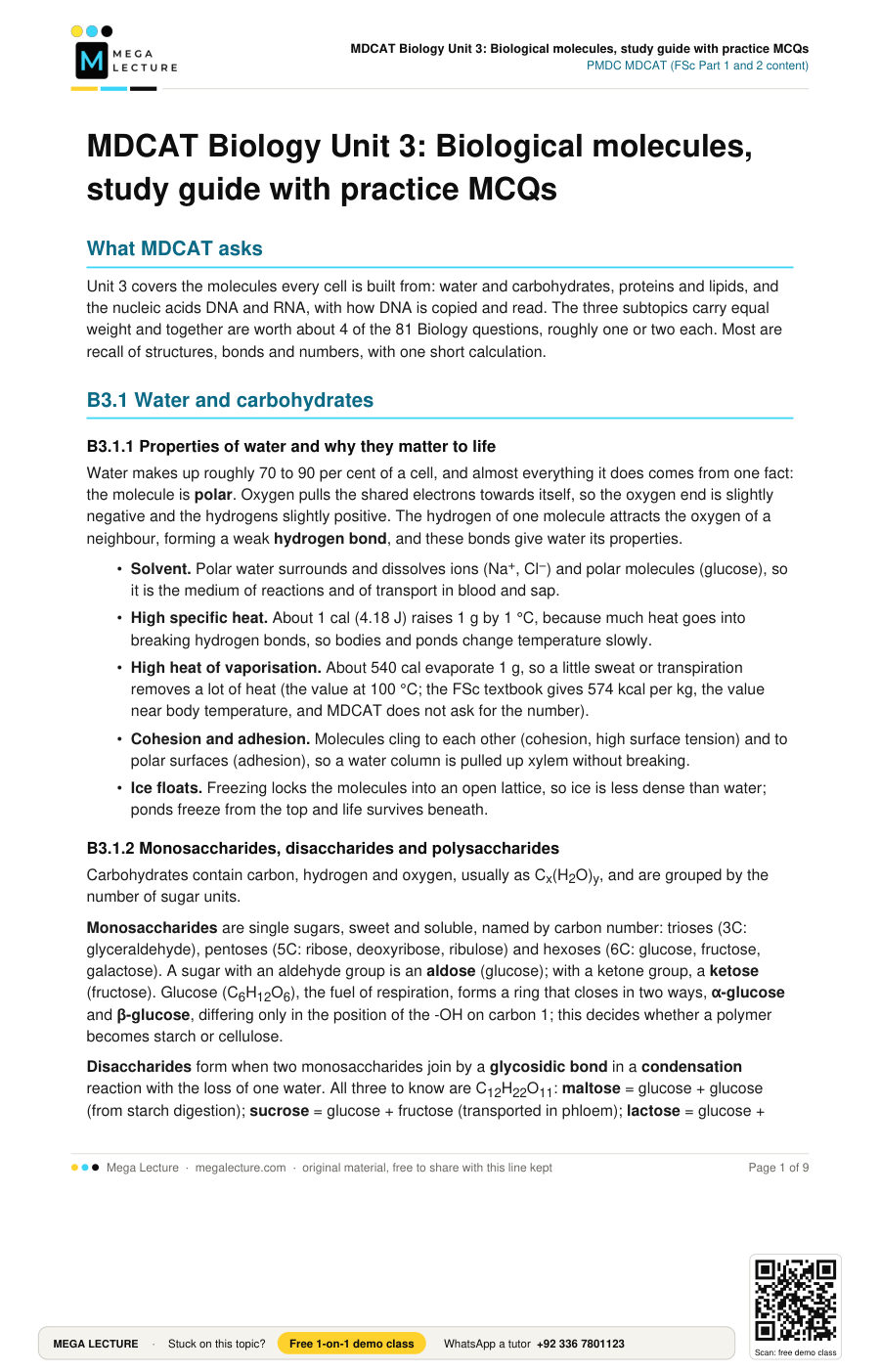
What MDCAT asks
Unit 3 covers the molecules every cell is built from: water and carbohydrates, proteins and lipids, and the nucleic acids DNA and RNA, with how DNA is copied and read. The three subtopics carry equal weight and together are worth about 4 of the 81 Biology questions, roughly one or two each. Most are recall of structures, bonds and numbers, with one short calculation.
B3.1 Water and carbohydrates
B3.1.1 Properties of water and why they matter to life
Water makes up roughly 70 to 90 per cent of a cell, and almost everything it does comes from one fact: the molecule is polar. Oxygen pulls the shared electrons towards itself, so the oxygen end is slightly negative and the hydrogens slightly positive. The hydrogen of one molecule attracts the oxygen of a neighbour, forming a weak hydrogen bond, and these bonds give water its properties.
- Solvent. Polar water surrounds and dissolves ions (Na+, Cl−) and polar molecules (glucose), so it is the medium of reactions and of transport in blood and sap.
- High specific heat. About 1 cal (4.18 J) raises 1 g by 1 °C, because much heat goes into breaking hydrogen bonds, so bodies and ponds change temperature slowly.
- High heat of vaporisation. About 540 cal evaporate 1 g, so a little sweat or transpiration removes a lot of heat (the value at 100 °C; the FSc textbook gives 574 kcal per kg, the value near body temperature, and MDCAT does not ask for the number).
- Cohesion and adhesion. Molecules cling to each other (cohesion, high surface tension) and to polar surfaces (adhesion), so a water column is pulled up xylem without breaking.
- Ice floats. Freezing locks the molecules into an open lattice, so ice is less dense than water; ponds freeze from the top and life survives beneath.
B3.1.2 Monosaccharides, disaccharides and polysaccharides
Carbohydrates contain carbon, hydrogen and oxygen, usually as Cx(H2O)y, and are grouped by the number of sugar units.
Monosaccharides are single sugars, sweet and soluble, named by carbon number: trioses (3C: glyceraldehyde), pentoses (5C: ribose, deoxyribose, ribulose) and hexoses (6C: glucose, fructose, galactose). A sugar with an aldehyde group is an aldose (glucose); with a ketone group, a ketose (fructose). Glucose (C6H12O6), the fuel of respiration, forms a ring that closes in two ways, α-glucose and β-glucose, differing only in the position of the -OH on carbon 1; this decides whether a polymer becomes starch or cellulose.
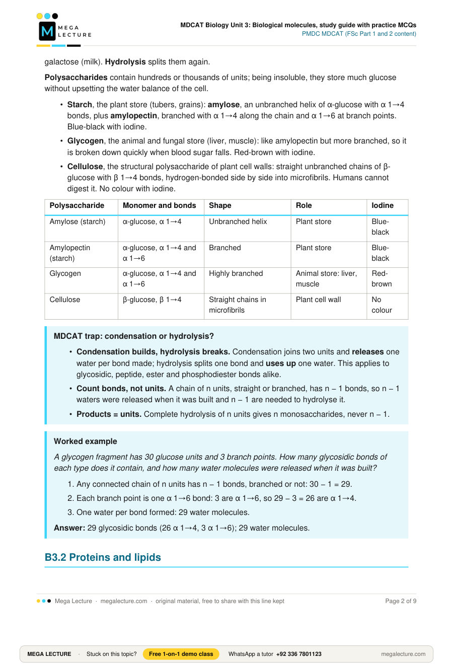
Disaccharides form when two monosaccharides join by a glycosidic bond in a condensation reaction with the loss of one water. All three to know are C12H22O11: maltose = glucose + glucose (from starch digestion); sucrose = glucose + fructose (transported in phloem); lactose = glucose + galactose (milk). Hydrolysis splits them again.
Polysaccharides contain hundreds or thousands of units; being insoluble, they store much glucose without upsetting the water balance of the cell.
- Starch, the plant store (tubers, grains): amylose, an unbranched helix of α-glucose with α 1→4 bonds, plus amylopectin, branched with α 1→4 along the chain and α 1→6 at branch points. Blue-black with iodine.
- Glycogen, the animal and fungal store (liver, muscle): like amylopectin but more branched, so it is broken down quickly when blood sugar falls. Red-brown with iodine.
- Cellulose, the structural polysaccharide of plant cell walls: straight unbranched chains of β-glucose with β 1→4 bonds, hydrogen-bonded side by side into microfibrils. Humans cannot digest it. No colour with iodine.
| Polysaccharide | Monomer and bonds | Shape | Role | Iodine |
|---|---|---|---|---|
| Amylose (starch) | α-glucose, α 1→4 | Unbranched helix | Plant store | Blue-black |
| Amylopectin (starch) | α-glucose, α 1→4 and α 1→6 | Branched | Plant store | Blue-black |
| Glycogen | α-glucose, α 1→4 and α 1→6 | Highly branched | Animal store: liver, muscle | Red-brown |
| Cellulose | β-glucose, β 1→4 | Straight chains in microfibrils | Plant cell wall | No colour |
MDCAT trap: condensation or hydrolysis?
- Condensation builds, hydrolysis breaks. Condensation joins two units and releases one water per bond made; hydrolysis splits one bond and uses up one water. This applies to glycosidic, peptide, ester and phosphodiester bonds alike.
- Count bonds, not units. A chain of n units, straight or branched, has n − 1 bonds, so n − 1 waters were released when it was built and n − 1 are needed to hydrolyse it.
- Products = units. Complete hydrolysis of n units gives n monosaccharides, never n − 1.
Worked example
A glycogen fragment has 30 glucose units and 3 branch points. How many glycosidic bonds of each type does it contain, and how many water molecules were released when it was built?
- Any connected chain of n units has n − 1 bonds, branched or not: 30 − 1 = 29.
- Each branch point is one α 1→6 bond: 3 are α 1→6, so 29 − 3 = 26 are α 1→4.
- One water per bond formed: 29 water molecules.
Answer: 29 glycosidic bonds (26 α 1→4, 3 α 1→6); 29 water molecules.
B3.2 Proteins and lipids
B3.2.1 Amino acids, the peptide bond and the levels of protein structure
Proteins are polymers of amino acids and contain carbon, hydrogen, oxygen and nitrogen, usually sulphur too. An amino acid has a central α-carbon carrying an amino group (-NH2), a carboxyl group (-COOH), a hydrogen atom and a variable R group. Only the R group differs (a single hydrogen in glycine, the simplest; a sulphur group in cysteine), and about 20 kinds build proteins.
The carboxyl group of one amino acid reacts with the amino group of the next; one water is removed and a peptide bond (-CO-NH-) forms. Many amino acids give a polypeptide with a free amino group at one end (N-terminus) and a free carboxyl at the other (C-terminus); a chain of n amino acids has n − 1 peptide bonds.
- Primary structure: the number and sequence of amino acids, held by peptide bonds and set by the gene.
- Secondary: regular coiling or folding held by hydrogen bonds between backbone C=O and N-H groups: the α-helix (keratin) or the β-pleated sheet (silk).
- Tertiary: the 3-D fold of the whole chain, held by bonds between R groups: hydrogen bonds, ionic bonds, disulphide bridges (covalent S-S links between two cysteines) and hydrophobic interactions.
- Quaternary: two or more chains held together by the same bonds. Haemoglobin has four chains (two α, two β), each with a haem group.
Fibrous proteins are long, insoluble and structural (collagen, keratin); globular proteins are compact, soluble and functional (enzymes, antibodies, haemoglobin). Heat, extreme pH and heavy metals break the weak bonds of the higher levels, so the protein loses shape and function (denaturation); peptide bonds survive, so the primary structure is unchanged.
B3.2.2 Triglycerides, phospholipids and other lipids
Lipids are not polymers; they are grouped together because they are insoluble in water but soluble in organic solvents.
Fatty acids are long hydrocarbon chains ending in a carboxyl group. A saturated fatty acid has only single C-C bonds, so the chain is straight and packs closely: fats rich in them (ghee, animal fat) are solid at room temperature. An unsaturated fatty acid has one or more C=C bonds, each putting a kink in the chain, so the molecules pack loosely and the lipid is a liquid oil.
Triglycerides (fats and oils) are the commonest lipids. One glycerol, a three-carbon alcohol with three -OH groups, condenses with three fatty acids; each -OH reacts with one -COOH to form an ester bond, so three ester bonds form and three water molecules are released. Triglycerides are the long-term energy store: fat gives roughly twice the energy per gram of carbohydrate.
Phospholipids are triglycerides in which one fatty acid is replaced by a phosphate group. The phosphate end is hydrophilic (the head); the two fatty acid chains are hydrophobic (the tails). In water the heads face outwards and the tails hide inside, forming the bilayer of every cell membrane.
Waxes are a fatty acid joined by an ester bond to a long-chain alcohol: waterproof coatings on leaves and insect cuticle. Steroids have four fused carbon rings; cholesterol sits in animal membranes and is the parent of the steroid hormones, vitamin D and bile salts. Terpenoids are built from five-carbon isoprene units: carotenoids (source of vitamin A), the phytol tail of chlorophyll, rubber.
| Lipid | Built from | Bonds | Main role |
|---|---|---|---|
| Triglyceride | 1 glycerol + 3 fatty acids | 3 ester bonds | Energy store, insulation, protection |
| Phospholipid | 1 glycerol + 2 fatty acids + phosphate | 2 ester bonds + phosphate link | Membrane bilayer |
| Wax | 1 fatty acid + 1 long-chain alcohol | 1 ester bond | Waterproofing |
| Steroid (cholesterol) | Four fused rings | No ester bond | Membranes, hormones, vitamin D, bile |
| Terpenoid (carotene) | Isoprene units | Carbon chain | Pigments, vitamin A, rubber |
MDCAT trap: counting peptide bonds and water molecules
The classic error is to say a protein of 100 amino acids has 100 peptide bonds. It has 99.
- One chain: bonds = amino acids − 1. Joining n units in a line needs n − 1 links; the two end residues each keep a free group.
- Several chains: subtract one per chain. A quaternary protein is several separate chains, each one bond short, so peptide bonds = total amino acids − number of chains. Chains are held together by hydrogen, ionic and disulphide bonds, never by peptide bonds.
- Water released = bonds formed, one per bond, not one per amino acid. Likewise a triglyceride forms three ester bonds and releases three waters.
Worked example
Insulin has an A chain of 21 amino acids and a B chain of 30. Two disulphide bridges join the chains and a third lies within the A chain. How many peptide bonds does one molecule contain, and what is the minimum number of cysteine residues?
- A chain: 21 − 1 = 20 peptide bonds; B chain: 30 − 1 = 29.
- The chains are joined by disulphide bridges, not peptide bonds, so the total is 20 + 29 = 49.
- Each disulphide bridge links two cysteines: 3 × 2 = 6 cysteines.
Answer: 49 peptide bonds; at least 6 cysteine residues.
B3.3 Nucleic acids
B3.3.1 The structure of DNA and RNA, and how they compare
Both DNA (deoxyribonucleic acid) and RNA (ribonucleic acid) are polymers of nucleotides. Every nucleotide has three parts: a pentose sugar (deoxyribose in DNA, ribose in RNA; deoxyribose lacks the oxygen on carbon 2′), a phosphate group on carbon 5′ of the sugar, and a nitrogenous base on carbon 1′.
Purines have two fused rings: adenine (A) and guanine (G). Pyrimidines have one ring: cytosine (C), thymine (T, DNA only) and uracil (U, RNA only).
Nucleotides join by condensation: the phosphate on carbon 5′ of one bonds to the -OH on carbon 3′ of the next, forming a phosphodiester bond. The chain has a sugar-phosphate backbone and a direction: a 5′ phosphate end and a 3′-OH end.
Watson and Crick (1953) showed that DNA is a double helix:
- Two strands wind around a common axis, backbones outside, bases inside like rungs of a ladder.
- The strands are antiparallel: one runs 5′ → 3′, the other 3′ → 5′.
- Complementary base pairing, always a purine with a pyrimidine: A with T by two hydrogen bonds, G with C by three. Every rung is the same width, so the helix is 2 nm across.
- Hence A = T and G = C, so purines equal pyrimidines (Chargaff's rule).
- One complete turn every 10 base pairs, 3.4 nm per turn, so adjacent base pairs are 0.34 nm apart.
RNA is usually a single strand and is made on a DNA template in the nucleus. Messenger RNA (mRNA) carries the code of one gene to the ribosome in triplets called codons; transfer RNA (tRNA), a clover-leaf molecule, has an anticodon that pairs with a codon and carries the matching amino acid; ribosomal RNA (rRNA) builds ribosomes.
| Feature | DNA | RNA |
|---|---|---|
| Sugar | Deoxyribose | Ribose |
| Bases | A, G, C, T | A, G, C, U |
| Strands | Two, antiparallel, double helix | One, may fold on itself |
| Pairing | A=T (2 H-bonds), G≡C (3 H-bonds) | A-U, G-C where it folds |
| Role | Stores hereditary information, self-replicates | Carries code (mRNA), carries amino acids (tRNA), builds ribosomes (rRNA) |
B3.3.2 DNA replication, genes, transcription and translation
Replication. Before a cell divides it copies its DNA semi-conservatively: each new helix keeps one old strand and gains one new strand.
- Helicase breaks the hydrogen bonds and separates the strands at a replication fork; the exposed bases act as templates.
- Primase lays down a short RNA primer, since DNA polymerase cannot start a strand.
- DNA polymerase III adds free nucleotides to the 3′ end of the new strand, so it grows only 5′ → 3′, pairing A with T and G with C against the template.
- The leading strand is made continuously towards the fork; the lagging strand is made backwards in short Okazaki fragments.
- DNA polymerase I replaces the RNA primers with DNA and DNA ligase seals the fragments.
A gene is a length of DNA carrying the instructions for one polypeptide. Information flows DNA → RNA → protein, the central dogma.
Transcription takes place in the nucleus. RNA polymerase binds the promoter at the start of the gene, unwinds it and uses one strand, the template (antisense) strand, to build mRNA 5′ → 3′, pairing A with U, T with A, G with C and C with G. The mRNA therefore has the same sequence as the other strand, the coding (sense) strand, with U in place of T.
The genetic code. mRNA is read in codons of three bases: 43 = 64 codons, of which 61 code for amino acids and 3 are stop codons (UAA, UAG, UGA). AUG is the start codon and codes for methionine.
Translation takes place on ribosomes in the cytoplasm. The ribosome binds the mRNA at AUG and moves one codon at a time; for each codon a tRNA with the complementary anticodon brings the right amino acid and a peptide bond joins it to the growing chain. At a stop codon no tRNA fits and the finished polypeptide is released.
MDCAT trap: which strand, and which base?
Two errors recur: putting T into an RNA sequence, and using the wrong DNA strand.
- RNA never contains thymine. If the question asks for mRNA, tRNA or rRNA and an option contains T, cross it out. DNA never contains U.
- mRNA is complementary to the template strand. Under each template base write its partner: A → U, T → A, G → C, C → G. mRNA is antiparallel to the template, so a template read 3′ → 5′ gives mRNA read 5′ → 3′.
- mRNA matches the coding strand. If you are given the coding (sense) strand, copy it and change every T to U.
Worked example
A double-stranded DNA molecule has 2,000 nucleotides, of which 20 per cent are thymine. Find the number of each base, the hydrogen bonds between the strands, the number of helix turns and the length.
- T = 20% of 2,000 = 400, so A = 400.
- The remaining 1,200 nucleotides are G and C in equal numbers: G = 600, C = 600.
- 1,000 base pairs: 400 A-T and 600 G-C. Hydrogen bonds = (400 × 2) + (600 × 3) = 2,600.
- One turn per 10 base pairs: 1,000 ÷ 10 = 100 turns.
- 0.34 nm per base pair: 1,000 × 0.34 = 340 nm.
Answer: A = 400, T = 400, G = 600, C = 600; 2,600 hydrogen bonds; 100 turns; 340 nm.
Practice MCQs
- Sweating cools the body because water has a:
A. high heat of vaporisation
B. high specific heat capacity
C. strong cohesion between molecules
D. lower density as ice than as liquid - Disaccharide X is hydrolysed to give only glucose. Disaccharide Y gives glucose and galactose. X and Y are, respectively:
A. sucrose and lactose
B. maltose and sucrose
C. maltose and lactose
D. lactose and maltose - An unbranched polysaccharide is digested completely to give 500 molecules of glucose. The number of water molecules used was:
A. 250
B. 500
C. 501
D. 499 - The α-helix of a polypeptide is held in shape mainly by:
A. hydrogen bonds between C=O and N-H groups of the backbone
B. peptide bonds between adjacent amino acids
C. disulphide bridges between cysteine residues
D. ionic bonds between charged R groups - The four chains of haemoglobin are separated but each keeps its own folded shape. The level of structure lost is the:
A. primary structure
B. quaternary structure
C. secondary structure
D. tertiary structure - A lipid is made from one glycerol and three fatty acids that contain no C=C double bonds. Which statement is correct?
A. Three waters released; liquid at room temperature
B. Two waters released; solid at room temperature
C. One water released; solid at room temperature
D. Three waters released; solid at room temperature - In a cell membrane, the part of each phospholipid that faces the watery cytoplasm is the:
A. two fatty acid tails
B. ester bonds
C. phosphate head
D. hydrocarbon chains - A nucleic acid extracted from a cell contains ribose and the base uracil. Which is also true of this molecule?
A. It contains thymine paired with adenine
B. It is usually a single strand
C. It is found only in the nucleus
D. It contains deoxyribose in every nucleotide - The template strand of a gene reads 3′-TAC GGA ATC-5′. The mRNA transcribed from it is:
A. 5′-AUG CCU UAG-3′
B. 5′-UAC GGA AUC-3′
C. 5′-ATG CCT TAG-3′
D. 5′-TAC GGA ATC-3′ - In a sample of double-stranded DNA, 30 per cent of the bases are adenine. The percentage of cytosine is:
A. 30
B. 70
C. 20
D. 40
Answer key
- A. Evaporating water takes a large amount of heat from the skin.
- C. Maltose is glucose + glucose; lactose is glucose + galactose. Sucrose would give glucose and fructose.
- D. A chain of 500 units has 499 glycosidic bonds, and hydrolysis uses one water per bond broken.
- A. Secondary structure is held by hydrogen bonds along the backbone. Peptide bonds give the primary structure.
- B. Quaternary structure is the association of two or more chains; each chain keeps its tertiary shape.
- D. Three fatty acids form three ester bonds with glycerol and release three waters. No C=C means saturated fatty acids, so the triglyceride is a solid fat, not an oil.
- C. The phosphate head is polar and hydrophilic, so it faces the water on both sides of the bilayer; the non-polar tails point inwards.
- B. Ribose and uracil mean RNA, which is usually single-stranded. It has no thymine or deoxyribose and works mostly in the cytoplasm.
- A. Pair each template base with its RNA partner (T → A, A → U, C → G, G → C): TAC → AUG, GGA → CCU, ATC → UAG, read 5′ → 3′.
- C. A = T, so A + T = 60 per cent; the remaining 40 per cent is G + C in equal amounts, so cytosine is 20 per cent.
Before the test: 12 facts to recite
- Water is polar; hydrogen bonds give its solvent action, high specific heat, high heat of vaporisation, cohesion and floating ice.
- Triose: glyceraldehyde; pentose: ribose, deoxyribose; hexose: glucose (aldose), fructose (ketose), galactose.
- Maltose = glucose + glucose; sucrose = glucose + fructose; lactose = glucose + galactose.
- Starch (amylose α 1→4, amylopectin branched α 1→6): plant store, blue-black with iodine. Glycogen: animal store, more branched, red-brown. Cellulose: β 1→4, straight, cell wall, no colour.
- Condensation joins units and releases water; hydrolysis breaks bonds and uses water; n units have n − 1 bonds.
- Amino acid: -NH2, -COOH, H and R group on one carbon; about 20 kinds; glycine simplest; peptide bond -CO-NH-.
- Primary = sequence (peptide bonds); secondary = α-helix or β-sheet (hydrogen bonds); tertiary = 3-D fold (hydrogen, ionic, disulphide, hydrophobic); quaternary = several chains (haemoglobin four).
- Triglyceride = glycerol + 3 fatty acids, 3 ester bonds, 3 waters; saturated (no C=C) solid fat, unsaturated oil; fat gives about twice the energy per gram of carbohydrate.
- Phospholipid: hydrophilic phosphate head, two hydrophobic tails, membrane bilayer. Cholesterol: four-ring steroid. Carotenoids: terpenoids.
- Nucleotide = pentose + phosphate + base; purines A, G (two rings); pyrimidines C, T, U (one ring); phosphodiester bonds link nucleotides.
- DNA: antiparallel double helix, A=T (2 hydrogen bonds), G≡C (3); 10 base pairs per turn, 3.4 nm per turn, 0.34 nm per pair, 2 nm wide. RNA: single strand, ribose, uracil.
- Replication: semi-conservative, helicase, primase, DNA polymerase 5′ → 3′, Okazaki fragments, ligase. Transcription: template strand → mRNA, U for T. Code: 64 codons, 61 sense, 3 stop (UAA, UAG, UGA), AUG start = methionine.
