
A Level · Biology 9700 · AS Biology 9700: full syllabus summary (all topics in one document)
AS Biology 9700: full syllabus summary (all topics in one document)
Opens inside this page. Prefer your own PDF app? Open the PDF.
Check yourself
5 quick questions on A Level Biology
Tap an answer to see if you are right, and why. Free, no sign-up.
Start the free test12 of 12 students found these notes useful
Was this note useful?
Full text of AS Biology 9700: full syllabus summary (all topics in one document)
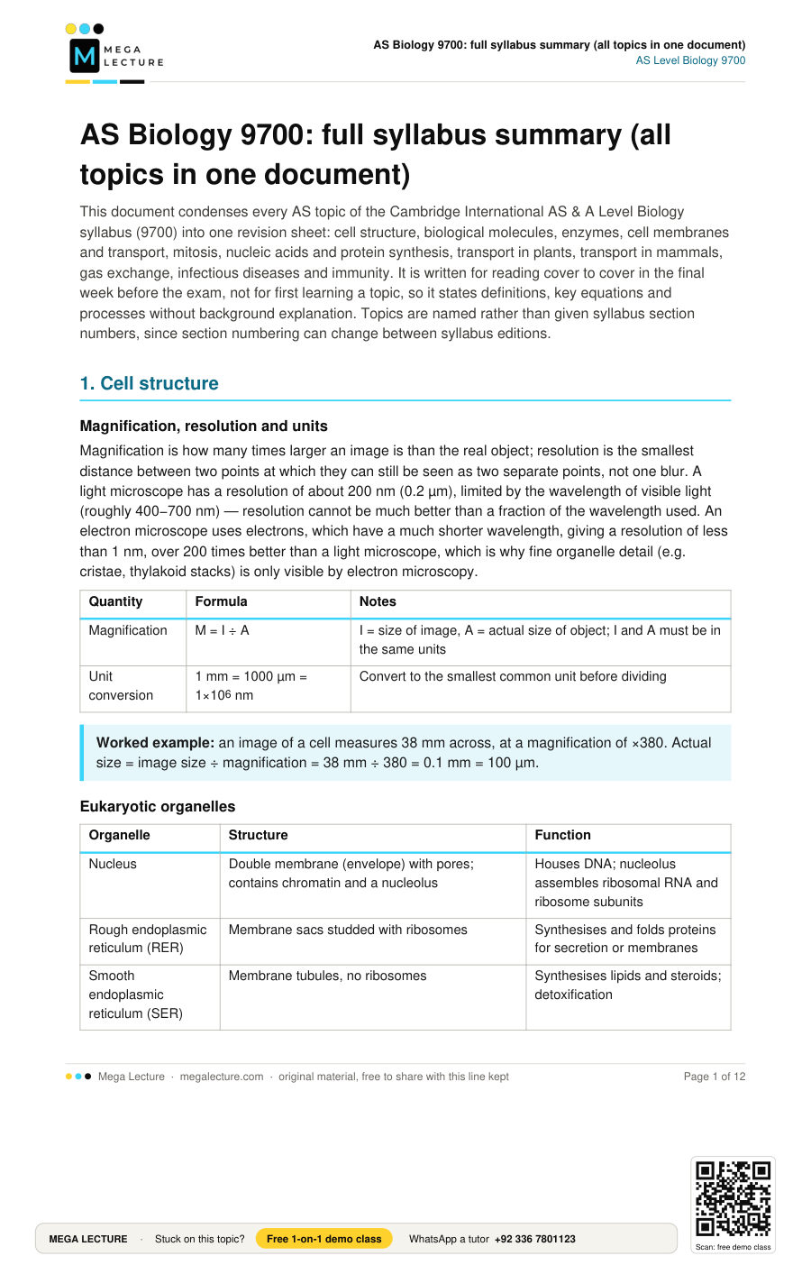
This document condenses every AS topic of the Cambridge International AS & A Level Biology syllabus (9700) into one revision sheet: cell structure, biological molecules, enzymes, cell membranes and transport, mitosis, nucleic acids and protein synthesis, transport in plants, transport in mammals, gas exchange, infectious diseases and immunity. It is written for reading cover to cover in the final week before the exam, not for first learning a topic, so it states definitions, key equations and processes without background explanation. Topics are named rather than given syllabus section numbers, since section numbering can change between syllabus editions.
1. Cell structure
Magnification, resolution and units
Magnification is how many times larger an image is than the real object; resolution is the smallest distance between two points at which they can still be seen as two separate points, not one blur. A light microscope has a resolution of about 200 nm (0.2 µm), limited by the wavelength of visible light (roughly 400−700 nm) — resolution cannot be much better than a fraction of the wavelength used. An electron microscope uses electrons, which have a much shorter wavelength, giving a resolution of less than 1 nm, over 200 times better than a light microscope, which is why fine organelle detail (e.g. cristae, thylakoid stacks) is only visible by electron microscopy.
| Quantity | Formula | Notes |
|---|---|---|
| Magnification | M = I ÷ A | I = size of image, A = actual size of object; I and A must be in the same units |
| Unit conversion | 1 mm = 1000 µm = 1×10⁶ nm | Convert to the smallest common unit before dividing |
Eukaryotic organelles
| Organelle | Structure | Function |
|---|---|---|
| Nucleus | Double membrane (envelope) with pores; contains chromatin and a nucleolus | Houses DNA; nucleolus assembles ribosomal RNA and ribosome subunits |
| Rough endoplasmic reticulum (RER) | Membrane sacs studded with ribosomes | Synthesises and folds proteins for secretion or membranes |
| Smooth endoplasmic reticulum (SER) | Membrane tubules, no ribosomes | Synthesises lipids and steroids; detoxification |
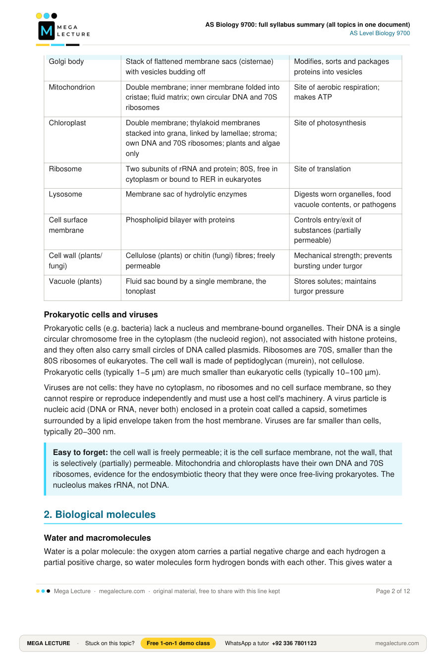
| Golgi body | Stack of flattened membrane sacs (cisternae) with vesicles budding off | Modifies, sorts and packages proteins into vesicles |
| Mitochondrion | Double membrane; inner membrane folded into cristae; fluid matrix; own circular DNA and 70S ribosomes | Site of aerobic respiration; makes ATP |
| Chloroplast | Double membrane; thylakoid membranes stacked into grana, linked by lamellae; stroma; own DNA and 70S ribosomes; plants and algae only | Site of photosynthesis |
| Ribosome | Two subunits of rRNA and protein; 80S, free in cytoplasm or bound to RER in eukaryotes | Site of translation |
| Lysosome | Membrane sac of hydrolytic enzymes | Digests worn organelles, food vacuole contents, or pathogens |
| Cell surface membrane | Phospholipid bilayer with proteins | Controls entry/exit of substances (partially permeable) |
| Cell wall (plants/fungi) | Cellulose (plants) or chitin (fungi) fibres; freely permeable | Mechanical strength; prevents bursting under turgor |
| Vacuole (plants) | Fluid sac bound by a single membrane, the tonoplast | Stores solutes; maintains turgor pressure |
Prokaryotic cells and viruses
Prokaryotic cells (e.g. bacteria) lack a nucleus and membrane-bound organelles. Their DNA is a single circular chromosome free in the cytoplasm (the nucleoid region), not associated with histone proteins, and they often also carry small circles of DNA called plasmids. Ribosomes are 70S, smaller than the 80S ribosomes of eukaryotes. The cell wall is made of peptidoglycan (murein), not cellulose. Prokaryotic cells (typically 1−5 µm) are much smaller than eukaryotic cells (typically 10−100 µm).
Viruses are not cells: they have no cytoplasm, no ribosomes and no cell surface membrane, so they cannot respire or reproduce independently and must use a host cell's machinery. A virus particle is nucleic acid (DNA or RNA, never both) enclosed in a protein coat called a capsid, sometimes surrounded by a lipid envelope taken from the host membrane. Viruses are far smaller than cells, typically 20−300 nm.
Easy to forget: the cell wall is freely permeable; it is the cell surface membrane, not the wall, that is selectively (partially) permeable. Mitochondria and chloroplasts have their own DNA and 70S ribosomes, evidence for the endosymbiotic theory that they were once free-living prokaryotes. The nucleolus makes rRNA, not DNA.2. Biological molecules
Water and macromolecules
Water is a polar molecule: the oxygen atom carries a partial negative charge and each hydrogen a partial positive charge, so water molecules form hydrogen bonds with each other. This gives water a high specific heat capacity (buffers temperature change), a high latent heat of vaporisation (evaporative cooling), cohesion between molecules (important in the transpiration stream) and makes it an excellent solvent for polar and ionic substances.
Large biological molecules (polysaccharides, proteins, nucleic acids) are polymers built from repeating monomer units. Monomers join by condensation reactions, forming a covalent bond and releasing one water molecule per bond; polymers are broken down by hydrolysis reactions, which use one water molecule to break a bond.
Carbohydrates
Monosaccharides (e.g. glucose, fructose, galactose) join by a condensation reaction between two −OH groups to form a glycosidic bond, releasing water. Two monosaccharides give a disaccharide: glucose + glucose → maltose; glucose + fructose → sucrose; glucose + galactose → lactose.
| Polysaccharide | Monomer/bonds | Structure and role |
|---|---|---|
| Amylose (part of starch) | α-glucose, 1,4 glycosidic bonds only | Unbranched, coils into a helix; compact storage in plants |
| Amylopectin (part of starch) | α-glucose, 1,4 and 1,6 glycosidic bonds | Branched; branches allow fast hydrolysis for rapid glucose release |
| Glycogen | α-glucose, 1,4 and 1,6 glycosidic bonds | More highly branched than amylopectin; storage in animals and fungi, very rapid glucose release |
| Cellulose | β-glucose, 1,4 glycosidic bonds; alternate glucose units rotated 180° | Long straight unbranched chains, H-bonded together into microfibrils; high tensile strength in plant cell walls |
Lipids
A triglyceride is one glycerol condensed with three fatty acids, forming three ester bonds. Unsaturated fatty acids contain one or more C=C double bonds, which kink the chain and lower melting point; saturated fatty acids have none. Phospholipids replace one fatty acid with a phosphate group, giving a hydrophilic head and two hydrophobic tails, the basis of the bilayer in cell membranes.
Proteins
An amino acid has a central carbon bonded to an amine group (−NH2), a carboxyl group (−COOH), a hydrogen, and a variable R group. Two amino acids condense to form a peptide bond between the carboxyl of one and the amine of the next.
| Level | What determines it |
|---|---|
| Primary | Sequence of amino acids joined by peptide bonds |
| Secondary | Hydrogen bonding between −NH and C=O groups of the backbone, forming an α-helix or β-pleated sheet |
| Tertiary | Further folding from R-group interactions: ionic bonds, hydrogen bonds, disulfide bonds and hydrophobic interactions |
| Quaternary | Two or more polypeptide chains associate (e.g. haemoglobin's four chains) |
Fibrous proteins have a structural rather than catalytic or transport role. Collagen, for example, is found in tendons, skin, bone and blood vessel walls: three polypeptide chains wind together into a triple helix, cross-linked by covalent bonds between adjacent molecules, giving it high tensile strength.
Biochemical tests
| Test | Method | Positive result |
|---|---|---|
| Reducing sugar | Add Benedict's reagent, heat in a water bath | Blue → green → yellow → brick-red precipitate |
| Non-reducing sugar | Boil with dilute HCl to hydrolyse, neutralise with alkali, then Benedict's test | Brick-red precipitate (only after hydrolysis) |
| Starch | Add iodine/potassium iodide solution | Blue-black colour |
| Protein | Biuret test: NaOH then dilute CuSO4 | Blue → purple/lilac |
| Lipid | Emulsion test: shake with ethanol, then add to water | White emulsion |
3. Enzymes
Enzyme action
Enzymes are globular proteins that act as biological catalysts: they speed up reactions by lowering the activation energy needed, without being used up. The induced-fit model describes how the active site is not a rigid fit but changes shape slightly as the substrate binds, moulding around it and straining bonds in the substrate to help the reaction proceed.
Some enzymes need a non-protein cofactor to function. An inorganic cofactor (e.g. Cl− or a metal ion such as Zn2+) assists catalysis without being part of the protein's structure. A coenzyme is a small organic molecule (e.g. NAD) that binds loosely and is often chemically changed during the reaction. A prosthetic group is a cofactor permanently and tightly bound to the enzyme (e.g. the haem group in catalase).
Factors affecting rate
| Factor | Effect |
|---|---|
| Temperature | Rate rises as kinetic energy and successful collisions increase (often roughly doubling per 10°C, the Q10 effect), up to an optimum; beyond the optimum, hydrogen and ionic bonds holding tertiary structure break, the active site changes shape (denaturation) and rate falls sharply |
| pH | Each enzyme has an optimum pH; above or below it, charges on R groups change, disrupting bonds that hold tertiary structure and denaturing the enzyme |
| Substrate concentration | Rate rises as concentration rises (more collisions), then plateaus at Vmax once all active sites are saturated |
| Enzyme concentration | Rate rises proportionally, provided substrate is in excess |
Inhibitors
| Type | Binding site | Effect of raising substrate concentration |
|---|---|---|
| Competitive | Active site (structurally resembles the substrate); reversible | Can outcompete the inhibitor, so Vmax can still be reached |
| Non-competitive | An allosteric site elsewhere on the enzyme, distorting the active site | Cannot be overcome; Vmax is reduced |
4. Cell membranes and transport
Fluid mosaic model
The membrane is a phospholipid bilayer with hydrophilic heads facing the aqueous cytoplasm and extracellular fluid, and hydrophobic tails facing each other. Proteins are scattered through it (a "mosaic") and can move within the layer (it is "fluid"): integral proteins span the bilayer (some acting as channels or carriers), peripheral proteins sit on one surface. Cholesterol sits between phospholipid tails and restricts their movement, regulating fluidity. Glycoproteins and glycolipids on the outer surface act as receptors and cell-recognition markers.
Movement across membranes
| Process | Direction | Uses ATP? | Route |
|---|---|---|---|
| Simple diffusion | High to low concentration | No | Directly through the bilayer (small, non-polar molecules, e.g. O2, CO2) |
| Facilitated diffusion | High to low concentration | No | Through a channel or carrier protein (large or polar/charged molecules, e.g. glucose, ions) |
| Osmosis | High to low water potential | No | Through the bilayer and water-channel proteins (aquaporins) |
| Active transport | Low to high concentration | Yes | Through a specific carrier protein that changes shape |
| Endocytosis / exocytosis | Bulk movement, in or out | Yes | Membrane folds around material to form a vesicle (endocytosis), or a vesicle fuses with the membrane to release its contents (exocytosis); used for large particles or bulk material rather than individual molecules |
Water potential (Ψ), measured in kPa, describes the tendency of water molecules to move out of a solution. Pure water has the highest possible water potential, 0 kPa; any solute lowers it to a negative value. Water always moves from a higher (less negative) to a lower (more negative) water potential.
In a plant cell, water potential is the sum of two components: Ψcell = Ψsolute + Ψpressure. Solute potential (Ψsolute) is always negative or zero, lowered by dissolved solutes; pressure potential (Ψpressure) is usually positive, produced by the protoplast pushing outward against the cell wall. In a hypotonic (dilute) solution, water enters the cell by osmosis and Ψpressure rises until it balances Ψsolute; the cell is then fully turgid and cannot burst, because the inelastic wall resists further expansion. In a hypertonic (concentrated) solution, water leaves the cell and the protoplast shrinks away from the wall (plasmolysis); at the point where the protoplast just starts to pull away from the wall, Ψpressure = 0 and Ψcell = Ψsolute (incipient plasmolysis).
Easy to forget: diffusion rate depends on surface area, concentration gradient and diffusion distance; link a structure's adaptations (thin, large surface area, short diffusion path) explicitly to these three factors.5. Mitosis
The cell cycle
Interphase, the longest phase, has three parts: G1 (growth, organelle production), S (DNA replication, so each chromosome goes from one to two sister chromatids) and G2 (further growth, checking DNA for errors). Mitosis then divides the nucleus, and cytokinesis divides the cytoplasm.
Stages of mitosis
| Stage | Key events |
|---|---|
| Prophase | Chromosomes condense and become visible, each as two sister chromatids joined at a centromere; the nuclear envelope breaks down; spindle fibres form |
| Metaphase | Chromosomes line up individually at the equator, attached to spindle fibres at the centromere |
| Anaphase | Centromeres split; sister chromatids are pulled to opposite poles by shortening spindle fibres |
| Telophase | Chromatids (now chromosomes) decondense; nuclear envelopes re-form around each set |
Mitosis produces two genetically identical daughter cells (same chromosome number and alleles as the parent), for growth, repair and asexual reproduction. Cancer results from mutations in genes controlling the cell cycle, causing uncontrolled mitotic division and tumour formation.
Stem cells
A stem cell is an undifferentiated cell that can divide by mitosis and differentiate into specialised cell types. Totipotent cells (e.g. the zygote) can become any cell type, including extra-embryonic tissue; pluripotent cells (e.g. embryonic stem cells) can become almost any body cell type, but not extra-embryonic tissue; multipotent cells (e.g. bone marrow stem cells) can become a limited range of related cell types. In plants, cells in the meristems at root and shoot tips remain undifferentiated and can divide throughout the plant's life, allowing continued growth.
Easy to forget: a chromosome is one DNA molecule; a chromatid is one half of a replicated chromosome. Chromosome number stays constant (diploid) throughout mitosis; only the chromatid number per chromosome changes across the cycle.6. Nucleic acids and protein synthesis
DNA structure and replication
A nucleotide is a phosphate group, a pentose sugar (deoxyribose in DNA, ribose in RNA) and a nitrogenous base. DNA is a double helix of two antiparallel strands, held together by hydrogen bonds between complementary bases: adenine pairs with thymine (2 hydrogen bonds), cytosine pairs with guanine (3 hydrogen bonds).
Replication is semi-conservative: helicase unwinds and unzips the double helix by breaking hydrogen bonds; DNA polymerase then builds a new complementary strand against each template strand, using free nucleotides, so each new DNA molecule keeps one original strand and one new strand.
| DNA | RNA | |
|---|---|---|
| Sugar | Deoxyribose | Ribose |
| Strands | Double | Single |
| Bases | A, T, C, G | A, U, C, G |
Protein synthesis
Transcription (in the nucleus): RNA polymerase binds upstream of a gene, unwinds the DNA and builds a single-stranded pre-mRNA copy of the template strand (using U in place of T). In eukaryotes, non-coding introns are then spliced out and the remaining exons joined to give mature mRNA, which leaves through a nuclear pore.
Translation (at ribosomes): mRNA is read three bases (a codon) at a time. Transfer RNA molecules, each carrying a specific amino acid and a complementary anticodon, pair with each codon in turn at the ribosome; the ribosome catalyses a peptide bond between adjacent amino acids, building a polypeptide until a stop codon is reached.
Easy to forget: the genetic code is a triplet code, non-overlapping and degenerate (most amino acids have more than one codon), but not ambiguous, since each codon specifies only one amino acid. AUG is both the start codon and codes for methionine.7. Transport in plants
Xylem and phloem
| Xylem | Phloem | |
|---|---|---|
| Cells | Vessel elements: dead, lignified, hollow tubes with no end walls, pits in the walls | Sieve tube elements (living, no nucleus, sieve plates with pores) and companion cells (living, nucleus, many mitochondria, linked by plasmodesmata) |
| Transports | Water and mineral ions | Organic solutes, mainly sucrose (translocation) |
| Direction | One way, roots to leaves | Source to sink, either direction depending on plant needs |
Transpiration
Water evaporates from moist mesophyll cell walls into the air spaces of the leaf and diffuses out through open stomata (the transpiration stream). This water loss lowers the water potential of the mesophyll cells, drawing water from the xylem by osmosis, which pulls the whole water column upward under tension. Cohesion between water molecules (hydrogen bonding) keeps this column continuous, and adhesion to xylem walls resists the column collapsing; this is the cohesion-tension theory. Root pressure (from active uptake of ions into the root xylem, lowering its water potential) gives an additional, smaller push. Transpiration rate increases with higher temperature, higher light intensity (more stomata open), lower humidity and higher wind speed (removes the water vapour boundary layer).
Translocation
The mass flow hypothesis: at the source (e.g. a leaf), companion cells actively load sucrose into sieve tubes, lowering the sieve tube's water potential, so water enters by osmosis from the xylem, raising hydrostatic pressure. At the sink (e.g. a root or fruit), sucrose is removed and used or stored, raising the sieve tube's water potential there, so water leaves by osmosis, lowering hydrostatic pressure. This pressure difference drives a mass (bulk) flow of phloem sap from source to sink.
Xerophytic adaptations
Plants in dry habitats (xerophytes) reduce water loss by transpiration in several ways: a thick waxy cuticle reduces evaporation directly through the leaf surface; sunken stomata, or stomata in pits or grooves, trap a layer of humid air next to the leaf and reduce the diffusion gradient; rolled leaves or leaf hairs also trap a boundary layer of moist air; a reduced leaf surface area, e.g. needle-shaped leaves, reduces the evaporating surface; and some plants shed leaves or close stomata during drought to cut water loss further.
Easy to forget: xylem transport is entirely passive (no ATP used); phloem loading at the source requires ATP for active transport of sucrose. Always use water-potential language ("higher/lower"), not "more/less water".8. Transport in mammals
The heart and cardiac cycle
Mammals have a double circulatory system: blood passes through the heart twice per full circuit, once to the lungs (pulmonary circulation) and once to the body (systemic circulation), keeping oxygenated and deoxygenated blood separate and maintaining high pressure. The heart has four chambers: right atrium and right ventricle handle deoxygenated blood; left atrium and left ventricle handle oxygenated blood. Atrioventricular valves (tricuspid on the right, bicuspid/mitral on the left) prevent backflow into the atria during ventricular contraction; semilunar valves in the pulmonary artery and aorta prevent backflow into the ventricles.
The heartbeat is myogenic: it is initiated by the sinoatrial node (SAN, the pacemaker) in the right atrium wall, which triggers atrial contraction (systole). The wave of excitation reaches the atrioventricular node (AVN), which imposes a short delay before conducting the impulse down the bundle of His and Purkyne fibres to the ventricle apex, so that ventricular systole spreads from the apex upward towards the base, squeezing blood upward and out through the semilunar valves.
Blood vessels and blood
| Artery | Vein | Capillary | |
|---|---|---|---|
| Wall | Thick, muscular and elastic | Thin | One cell thick (endothelium only) |
| Lumen | Narrow | Wide | Very narrow, single blood cell width |
| Valves | Absent (except at heart exits) | Present | Absent |
| Pressure | High, pulsing | Low, steady | Falls along its length |
Red blood cells are biconcave (large surface area to volume ratio for gas exchange) and packed with haemoglobin, a protein with quaternary structure: four polypeptide chains, each with a haem group containing Fe2+, each reversibly binding one O2, summarised as Hb + 4O2 ⇌ Hb(O2)4. Binding of the first O2 makes further binding easier (cooperative binding), producing an S-shaped (sigmoid) oxygen dissociation curve. A rise in CO2 concentration (and fall in pH) shifts the curve to the right, the Bohr effect, reducing haemoglobin's affinity for O2 so more is released at respiring tissues.
Tissue fluid
At the arterial end of a capillary bed, high hydrostatic pressure forces plasma (minus large proteins) out through gaps in the capillary wall into the surrounding tissue, forming tissue fluid. At the venous end, hydrostatic pressure has fallen while the plasma proteins left behind lower the water potential inside the capillary, so most tissue fluid re-enters by osmosis; the small remaining excess is drained by the lymphatic system and returned to the blood.
Easy to forget: fetal haemoglobin has a higher affinity for O2 than adult haemoglobin (its dissociation curve sits to the left), allowing O2 to transfer from mother to fetus across the placenta.9. Gas exchange
Structure of the gas exchange system
Air passes through the trachea (supported by C-shaped cartilage rings, keeping it open), which branches into two bronchi, then into narrower bronchioles, ending in clusters of alveoli. Alveoli provide the exchange surface: their walls are a single layer of squamous epithelium, they are surrounded by a dense capillary network with walls also one cell thick (a short diffusion distance), there are millions of them (a very large total surface area, around 70 m2 in humans), and their inner surface is moist, allowing gases to dissolve before diffusing.
Ventilation
| Inspiration | Expiration (quiet breathing) | |
|---|---|---|
| External intercostal muscles | Contract, pulling ribs up and out | Relax |
| Diaphragm | Contracts, flattens | Relaxes, domes upward |
| Thoracic volume | Increases | Decreases |
| Pressure inside thorax | Falls below atmospheric | Rises above atmospheric |
| Airflow | Into the lungs | Out of the lungs |
10. Infectious diseases
Pathogens and transmission
Pathogens causing disease belong to several groups: bacteria, viruses, protoctists and fungi. Transmission can be direct (e.g. droplet infection, direct contact, sexual contact) or indirect (e.g. via contaminated water or food, or via a vector such as a mosquito).
| Disease | Pathogen and type | Transmission / key mechanism |
|---|---|---|
| Cholera | Vibrio cholerae, bacterium | Faecal-contaminated water; a toxin causes chloride ion secretion into the gut lumen, water follows by osmosis, causing severe watery diarrhoea and dehydration |
| Tuberculosis (TB) | Mycobacterium tuberculosis, bacterium | Airborne droplets; infects the lungs, forming areas of damaged tissue (tubercles) |
| Malaria | Plasmodium species, protoctist | Vector-borne, transmitted by Anopheles mosquitoes; the parasite develops in the liver, then repeatedly infects and bursts red blood cells |
| HIV/AIDS | HIV, a virus | Transmitted via bodily fluids (blood, semen, breast milk); infects and destroys helper T cells, eventually collapsing immune function (AIDS) |
| Measles | Measles virus | Airborne droplets; highly infectious |
Antibiotics
Antibiotics kill or inhibit bacteria without harming host cells by targeting structures or processes unique to bacteria: for example, penicillin-type antibiotics inhibit the enzyme that cross-links peptidoglycan in the bacterial cell wall, so the wall weakens and the bacterium lyses (bursts) as it grows. Antibiotics are ineffective against viruses, which have no cell wall, ribosomes or independent metabolism to target. Antibiotic resistance arises when a random mutation gives some bacteria a survival advantage in the presence of the antibiotic; these survive, reproduce and pass the resistance allele on, so the resistant strain increases in frequency by natural selection.
Easy to forget: antibiotics fail against colds or flu because those are caused by viruses, not because the antibiotic is "too weak"; always explain in terms of the pathogen type and the antibiotic's target.11. Immunity
Non-specific defences
Physical and chemical barriers (skin, mucous membranes, stomach acid, lysozyme enzyme in tears and saliva) prevent pathogens entering. If a pathogen breaches these, phagocytes (e.g. macrophages, neutrophils) engulf it by phagocytosis: the pathogen is enclosed in a phagosome, which fuses with a lysosome, and hydrolytic enzymes digest it. This response is the same regardless of the pathogen (non-specific).
Specific immune response
Each pathogen carries surface molecules called antigens, recognised by lymphocytes with a complementary receptor. Humoral immunity: a B lymphocyte with a matching receptor binds the antigen, is activated (with help from a helper T cell) and undergoes clonal selection and division, producing plasma cells that secrete large quantities of antibodies specific to that antigen, and memory cells that persist. Cell-mediated immunity: helper T cells recognise antigen presented on an infected cell's surface and release cytokines that activate B cells and cytotoxic T cells; cytotoxic T cells then destroy infected body cells directly.
An antibody is a protein with quaternary structure: two identical heavy chains and two identical light chains, joined by disulfide bonds, forming a Y-shape with two identical variable regions (antigen-binding sites, complementary to one specific antigen) and a constant region.
Primary and secondary response; types of immunity
On first exposure, the primary response is slow to reach a high antibody concentration because clonal selection of the rare matching lymphocyte takes time; the pathogen may cause disease symptoms before it is cleared. Memory cells remain afterwards. On re-exposure, the secondary response is faster and reaches a much higher antibody concentration, because memory cells recognise the antigen immediately and divide rapidly, often clearing the pathogen before symptoms appear.
| Active immunity | Passive immunity | |
|---|---|---|
| Source of antibodies | Made by the individual's own plasma cells | Given from an outside source |
| Memory cells formed? | Yes | No |
| Duration | Long-lasting | Short-term |
| Natural example | Recovering from infection | Antibodies crossing the placenta / in breast milk |
| Artificial example | Vaccination | Injection of antibodies (antiserum) |
Vaccination introduces antigens (from a dead or attenuated pathogen, a toxoid, or an isolated antigen) to trigger a primary response and memory cell formation without causing disease, so a real infection later triggers a fast, protective secondary response. If enough of a population is vaccinated, the pathogen cannot spread easily even to unvaccinated individuals, giving herd immunity.
Easy to forget: antibodies do not destroy pathogens directly; they mark them for destruction (agglutination, or as a signal for phagocytes) and neutralise toxins or prevent attachment to host cells.Common mistakes
- Writing "the enzyme is destroyed" instead of "denatured": the bonds maintaining tertiary structure break, but peptide bonds and primary structure are unaffected.
- Confusing diffusion and osmosis: osmosis is specifically the movement of water across a partially permeable membrane down a water potential gradient; do not use "concentration gradient" language for osmosis in mark schemes.
- Stating water potential is "high" or "low" without a comparison: always compare two named regions, e.g. "water moves from the cell (higher Ψ) to the solution (lower Ψ)".
- Mixing up chromosome and chromatid, or the chromosome-number change in mitosis: mitosis keeps the chromosome number constant; it is the chromatid number that changes through the cycle.
- Treating translocation and transpiration as the same process: transpiration is passive water movement in xylem; translocation is active loading followed by mass flow of solutes in phloem.
- Omitting units: always state units for water potential (kPa), rate calculations (e.g. cm3 min−1) and magnification (a ratio, so no unit).
- Assuming antibiotics work on viral infections: viruses lack the bacterial structures, such as a peptidoglycan cell wall, that antibiotics target.
- Confusing an antigen with an antibody: an antigen triggers the immune response; an antibody is produced in response to it.
- Describing a structure without linking it to function: for "describe" or "explain how X is adapted" questions, always pair the feature with its functional consequence.
