
A Level · Biology 9700 · Immunity: summary notes
Immunity: summary notes
Opens inside this page. Prefer your own PDF app? Open the PDF.
Check yourself
5 quick questions on A Level Biology
Tap an answer to see if you are right, and why. Free, no sign-up.
Start the free test12 of 12 students found these notes useful
Was this note useful?
Full text of Immunity: summary notes
These notes cover Topic 11 (Immunity) of the Cambridge International AS & A Level Biology syllabus (9700). They start with the non-specific response of phagocytes and then cover the specific response of B and T lymphocytes, primary and secondary responses, antibody structure and function, types of immunity, vaccination, autoimmune disease and monoclonal antibodies. Each section gives the key facts. Boxes hold the definitions you should learn word for word and the worked examples. After Section 8 there is a list of common exam errors. A quick-check set of questions with answers is at the end. Material labelled "Extension" goes beyond the syllabus learning outcomes. It is useful background, but you will not be asked about it directly.
1. Phagocytes and phagocytosis
Syllabus reference: 11.1
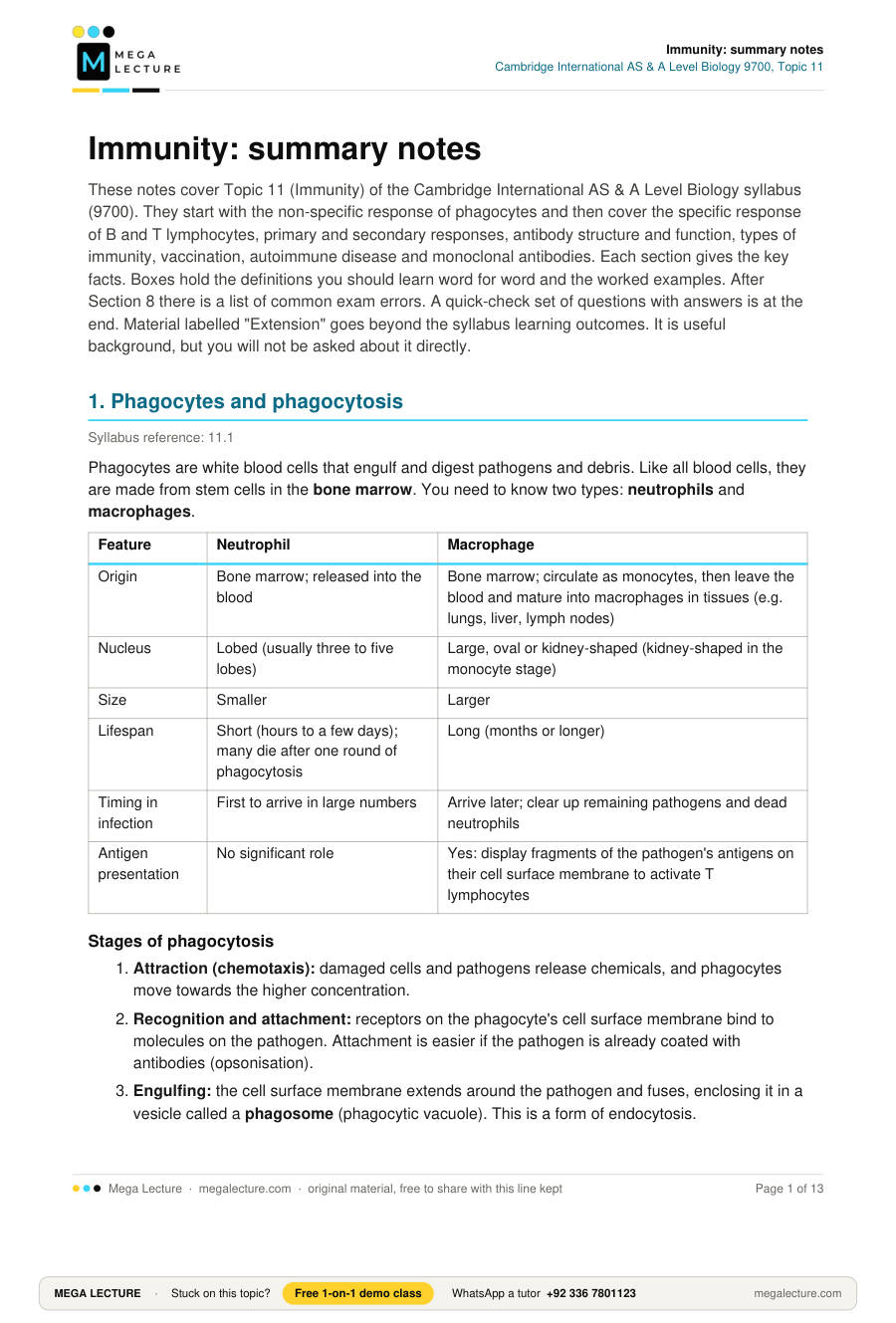
Phagocytes are white blood cells that engulf and digest pathogens and debris. Like all blood cells, they are made from stem cells in the bone marrow. You need to know two types: neutrophils and macrophages.
| Feature | Neutrophil | Macrophage |
|---|---|---|
| Origin | Bone marrow; released into the blood | Bone marrow; circulate as monocytes, then leave the blood and mature into macrophages in tissues (e.g. lungs, liver, lymph nodes) |
| Nucleus | Lobed (usually three to five lobes) | Large, oval or kidney-shaped (kidney-shaped in the monocyte stage) |
| Size | Smaller | Larger |
| Lifespan | Short (hours to a few days); many die after one round of phagocytosis | Long (months or longer) |
| Timing in infection | First to arrive in large numbers | Arrive later; clear up remaining pathogens and dead neutrophils |
| Antigen presentation | No significant role | Yes: display fragments of the pathogen's antigens on their cell surface membrane to activate T lymphocytes |
Stages of phagocytosis
- Attraction (chemotaxis): damaged cells and pathogens release chemicals, and phagocytes move towards the higher concentration.
- Recognition and attachment: receptors on the phagocyte's cell surface membrane bind to molecules on the pathogen. Attachment is easier if the pathogen is already coated with antibodies (opsonisation).
- Engulfing: the cell surface membrane extends around the pathogen and fuses, enclosing it in a vesicle called a phagosome (phagocytic vacuole). This is a form of endocytosis.
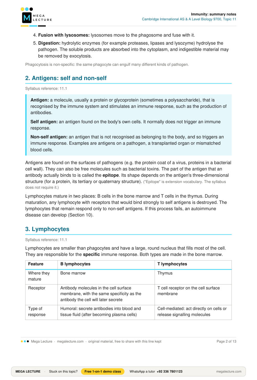
- Fusion with lysosomes: lysosomes move to the phagosome and fuse with it.
- Digestion: hydrolytic enzymes (for example proteases, lipases and lysozyme) hydrolyse the pathogen. The soluble products are absorbed into the cytoplasm, and indigestible material may be removed by exocytosis.
Phagocytosis is non-specific: the same phagocyte can engulf many different kinds of pathogen.
2. Antigens: self and non-self
Syllabus reference: 11.1
Antigen: a molecule, usually a protein or glycoprotein (sometimes a polysaccharide), that is recognised by the immune system and stimulates an immune response, such as the production of antibodies.
Self antigen: an antigen found on the body's own cells. It normally does not trigger an immune response.
Non-self antigen: an antigen that is not recognised as belonging to the body, and so triggers an immune response. Examples are antigens on a pathogen, a transplanted organ or mismatched blood cells.
Antigens are found on the surfaces of pathogens (e.g. the protein coat of a virus, proteins in a bacterial cell wall). They can also be free molecules such as bacterial toxins. The part of the antigen that an antibody actually binds to is called the epitope. Its shape depends on the antigen's three-dimensional structure (for a protein, its tertiary or quaternary structure). ("Epitope" is extension vocabulary. The syllabus does not require it.)
Lymphocytes mature in two places: B cells in the bone marrow and T cells in the thymus. During maturation, any lymphocyte with receptors that would bind strongly to self antigens is destroyed. The lymphocytes that remain respond only to non-self antigens. If this process fails, an autoimmune disease can develop (Section 10).
3. Lymphocytes
Syllabus reference: 11.1
Lymphocytes are smaller than phagocytes and have a large, round nucleus that fills most of the cell. They are responsible for the specific immune response. Both types are made in the bone marrow.
| Feature | B lymphocytes | T lymphocytes |
|---|---|---|
| Where they mature | Bone marrow | Thymus |
| Receptor | Antibody molecules in the cell surface membrane, with the same specificity as the antibody the cell will later secrete | T cell receptor on the cell surface membrane |
| Type of response | Humoral: secrete antibodies into blood and tissue fluid (after becoming plasma cells) | Cell-mediated: act directly on cells or release signalling molecules |
| Cell types produced | Plasma cells, memory B cells | T helper cells, T killer cells, memory T cells |
| Antigens recognised | Free antigens and antigens on pathogens | Antigens presented on the surface of body cells (e.g. macrophages or infected cells) |
Each lymphocyte has receptors of only one shape, so each can respond to only one antigen. There are millions of different lymphocytes, each with a different receptor, so almost any antigen will be complementary to the receptors of a few lymphocytes.
4. B lymphocytes, plasma cells and memory cells
Syllabus reference: 11.1
- Clonal selection: a B lymphocyte whose receptors are complementary in shape to a particular antigen binds to it. Only this B cell (and others with the same receptor) is selected.
- Activation: T helper cells that recognise the same antigen release cytokines, which stimulate the B cell further.
- Clonal expansion: the selected B cell divides repeatedly by mitosis, producing a clone of genetically identical cells.
- Differentiation: most cells of the clone become plasma cells. Some become memory B cells.
Plasma cells are short-lived (days to weeks). Each secretes large quantities of one type of antibody, specific to the antigen that triggered the response. They are adapted for this in four ways:
- extensive rough endoplasmic reticulum, for protein synthesis;
- a large Golgi body, which modifies and packages the antibody;
- many mitochondria, which supply ATP for synthesis and exocytosis;
- many secretory vesicles.
Memory B cells are long-lived and stay in the blood and lymph nodes. They do not secrete antibodies. If the same antigen is met again, they divide rapidly to form plasma cells.
Memory cell: a long-lived lymphocyte, produced during a primary immune response, that stays in the body. If the same antigen enters the body again, it responds rapidly by dividing. Memory cells are the basis of long-term immunity.
5. T helper and T killer cells
Syllabus reference: 11.1
T lymphocytes only recognise an antigen when it is presented on the surface of a body cell. This body cell may be a macrophage that has digested a pathogen (an antigen-presenting cell) or a body cell infected with a virus. A T cell with a complementary receptor binds to the antigen, is selected and divides by mitosis (clonal expansion). The clone differentiates into three cell types:
| Cell | What it does |
|---|---|
| T helper cell | Releases cytokines (signalling molecules, e.g. interleukins). These have three effects: they stimulate the matching B cells to divide and become plasma cells, stimulate macrophages to carry out more phagocytosis, and stimulate T killer cells to divide. |
| T killer cell (cytotoxic T cell) | Binds to body cells showing the specific non-self antigen (infected cells or some cancer cells). It then releases toxic substances that kill the cell: perforin, which makes pores in the target cell's membrane, and enzymes that make the cell die. Killing the infected cell stops the virus from replicating inside it and from releasing new virus particles to infect other cells. |
| Memory T cell | Long-lived; divides rapidly on a second exposure to the same antigen. |
Remember: T killer cells kill infected body cells, not the free pathogen. Antibodies and phagocytes deal with free pathogens.
Worked example 1: putting the sequence together
Question. A virus enters the body for the first time. Outline the sequence of events leading to the production of antibodies against it. [5]
Step 1: identify the start. The virus is non-self, so a macrophage engulfs it by phagocytosis and digests it.
Step 2: presentation. The macrophage displays viral antigens on its cell surface membrane (antigen presentation).
Step 3: T helper activation. A T helper cell with a complementary receptor binds, is selected and divides by mitosis. Its clone releases cytokines.
Step 4: B cell selection. A B cell with receptors complementary to the viral antigen binds to it, and cytokines from the T helper cells stimulate it.
Step 5: expansion and differentiation. The B cell divides by mitosis (clonal expansion). The clone differentiates into plasma cells, which secrete specific antibodies, and memory B cells.
A mark scheme for this question would list more creditable points than the 5 marks available: phagocytosis, antigen presentation, T helper selection, cytokines, B cell clonal selection, mitosis, plasma cells, antibody secretion and memory cells. Write each point as a separate precise statement, in order, using the technical terms (clonal selection, clonal expansion, mitosis, plasma cell, cytokines).
6. Primary and secondary immune responses
Syllabus reference: 11.1
The primary response happens the first time an antigen is met. There is a delay while the few lymphocytes with complementary receptors are found, activated and cloned. Antibody concentration therefore rises slowly and reaches a relatively low peak. During this time the person may develop symptoms. Memory cells are produced.
The secondary response happens when the same antigen is met again. There are now many memory cells, so the antigen is recognised quickly and the memory cells divide rapidly into plasma cells. The response is faster, larger and lasts longer. The pathogen is usually destroyed before symptoms appear, so the person is immune.
The classic graph (described in words)
The x-axis is time after first exposure (days). The y-axis is antibody concentration in the blood in arbitrary units, often on a log scale.
- After the first exposure (day 0), the line stays near zero for several days (the lag phase). It then rises gradually to a small peak and falls again.
- The second exposure, weeks or months later, is marked by an arrow on the x-axis. The line rises almost immediately and very steeply to a peak many times higher than the first, and it falls much more slowly.
| Feature | Primary response | Secondary response |
|---|---|---|
| Lag before antibodies appear | Longer (several days) | Shorter (a day or two) |
| Rate of antibody production | Slower | Faster |
| Peak concentration | Lower | Much higher |
| Duration of high antibody level | Short | Longer |
| Cells responding | Few naive B and T cells | Many memory cells |
| Symptoms | Often present | Usually none |
Worked example 2: data from a response curve
A student measured the antibody concentration in a rabbit that was given the same harmless antigen twice. The data are invented.
| Event | Days after that exposure until peak | Peak concentration / arbitrary units |
|---|---|---|
| First exposure | 12 | 16 |
| Second exposure (day 40) | 5 | 240 |
(a) Calculate how many times greater the secondary peak is than the primary peak. [1]
Ratio = 240 ÷ 16 = 15 (the secondary peak is 15 times greater).
(b) Calculate the percentage increase in peak concentration. [2]
Increase = 240 − 16 = 224 arbitrary units.
Percentage increase = (224 ÷ 16) × 100 = 1400%.
(c) Calculate the mean rate of increase in antibody concentration from exposure to peak for each response. Assume the concentration was 0 at each exposure. [2]
Primary: 16 ÷ 12 = 1.3 arbitrary units per day (to 2 s.f.).
Secondary: 240 ÷ 5 = 48 arbitrary units per day.
(d) Explain the difference. [3]
Memory B cells were produced in the primary response; there are many more of them than the original naive B cells; they recognise the antigen quickly and divide rapidly by mitosis into plasma cells, so antibodies are secreted sooner and faster.
Note: the assumption in (c) that the concentration was zero at the second exposure is a simplification. In reality, some antibody from the first response may remain.
7. Antibody structure and function
Syllabus reference: 11.2
Antibody: a globular glycoprotein (an immunoglobulin), secreted by plasma cells, that binds specifically to a particular antigen.
Structure
A typical antibody (IgG) is Y-shaped. It is made of four polypeptide chains, so it has quaternary structure:
- Two heavy chains (long) and two light chains (short). The two heavy chains are identical, and so are the two light chains.
- Disulfide bonds hold the chains together: between each light chain and its heavy chain, and between the two heavy chains.
- Variable regions are at the tips of both arms. Each is formed by parts of one heavy chain and one light chain. Their amino acid sequence differs between antibodies, which gives each antibody a unique antigen-binding site complementary in shape to one specific antigen. Each antibody has two identical antigen-binding sites.
- Constant regions make up the rest of the molecule. Their sequence is the same for all antibodies of one class. The constant region of the stem binds to receptors on phagocytes and can activate other parts of the immune system.
- The hinge region is where the arms meet the stem. It is flexible, so the distance and angle between the two binding sites can vary. One antibody can therefore bind to two antigens on different pathogens, or to two antigens spaced differently on the same pathogen.
- Carbohydrate chains are attached to the heavy chains, which is why an antibody is a glycoprotein.
Diagram in words: draw a Y. Each arm is one light chain lying alongside the upper part of a heavy chain. The tips of the arms are the variable regions. The stem is formed by the lower parts of the two heavy chains. Label the disulfide bonds across the hinge and between the light and heavy chains.
Functions (relating structure to function)
| Function | How it works | Structural feature involved |
|---|---|---|
| Agglutination | Each antibody binds to antigens on two pathogens, so the pathogens clump together. This stops them spreading and makes it easier for phagocytes to engulf many pathogens at once. | Two binding sites; flexible hinge |
| Neutralisation | Antibodies bind to toxins and block the toxin's active part, so it cannot harm cells. In this role they are called antitoxins. | Specific variable region |
| Blocking attachment | Antibodies bind to the binding sites on viruses or bacteria, so the pathogens cannot attach to or enter host cells. | Specific variable region |
| Opsonisation (marking for phagocytes) | Antibodies coat the pathogen. Phagocytes have receptors for the constant region, so they bind to and engulf the pathogen more readily. | Constant region |
| Activating complement / lysis (Extension) | Bound antibodies trigger blood proteins that punch holes in bacterial cell membranes, causing lysis. | Constant region |
8. Types of immunity
Syllabus reference: 11.2
Active immunity: immunity gained when an antigen stimulates the body's own lymphocytes to produce antibodies and memory cells. It takes time to develop but is long-lasting.
Passive immunity: immunity gained when antibodies made by another organism are introduced into the body. It is immediate but short-lived, because no memory cells are made and the antibodies are gradually broken down.
Natural immunity arises through normal life (infection, or from mother to child). Artificial immunity arises through medical intervention (vaccination or injection of antibodies).
| Natural | Artificial | |
|---|---|---|
| Active | Catching an infection, e.g. recovering from chickenpox | Vaccination, e.g. measles vaccine |
| Passive | Antibodies crossing the placenta to the fetus; antibodies in breast milk, especially colostrum | Injection of ready-made antibodies, e.g. antitoxin for tetanus or antivenom after a snake bite |
| Feature | Active | Passive |
|---|---|---|
| Antibodies made by | The person's own plasma cells | Another person or animal |
| Memory cells produced? | Yes | No |
| Speed of protection | Delayed (lag of days to weeks) | Immediate |
| Duration | Long-term (years, sometimes life) | Short-term (weeks to months) |
Common exam errors
Each line gives a common mistake, then the correct version.
- Mixing up antigen and antibody. The antigen is the molecule that triggers the response (e.g. on the pathogen). The antibody is the protein made by plasma cells that binds to it.
- "Antibodies kill pathogens." Antibodies do not kill pathogens directly. They cause agglutination, neutralisation or opsonisation, and then phagocytes destroy the pathogen.
- "Memory cells secrete antibodies." Memory cells do not secrete antibodies. They divide to form plasma cells, and the plasma cells secrete antibodies.
- "Vaccines contain antibodies." Vaccines contain antigens (or the instructions to make them). Injections of ready-made antibodies give passive immunity and are not vaccines.
- "Same shape". Write "complementary shape". An antibody's binding site is complementary to the antigen, not the same shape as it.
- "Plasma cells divide." Plasma cells do not divide. The selected B cell divides by mitosis, and its clone then differentiates into plasma cells.
- "T killer cells engulf pathogens." T killer cells do not engulf anything. They bind to infected body cells and kill them with perforin and enzymes. Engulfing is done by phagocytes.
- "Passive immunity produces memory cells." Passive immunity produces no memory cells, which is why it is short-lived.
9. Vaccination and herd immunity
Syllabus reference: 11.2
Vaccine: a preparation containing antigens (from a pathogen, or instructions to make them) that is given to stimulate a primary immune response. Memory cells are made without the person suffering the disease.
Vaccines may contain:
- live but weakened (attenuated) pathogens, e.g. measles, BCG for TB;
- killed (inactivated) pathogens, e.g. some polio vaccines;
- modified toxins (toxoids), e.g. tetanus;
- isolated antigens (subunit vaccines), e.g. hepatitis B surface protein;
- genetic material coding for an antigen, e.g. mRNA vaccines against COVID-19.
Booster doses cause a secondary response, which raises the number of memory cells and the antibody level.
Herd immunity
When a large enough proportion of a population is immune, the pathogen has too few susceptible hosts to pass between, so chains of transmission break. This protects people who cannot be vaccinated, such as newborns, people with weak immune systems and people allergic to vaccine components. The more infectious the disease, the higher the percentage of the population that must be vaccinated.
Why some diseases are hard to control by vaccination
- Antigenic variation: the pathogen's antigens change (e.g. influenza virus, HIV), so memory cells no longer recognise it.
- Several strains or many antigens: e.g. Plasmodium, which also hides inside liver cells and red blood cells.
- Intracellular pathogens are shielded from antibodies.
- Practical problems: cold storage, remote communities, refugees moving between areas, public distrust.
Extension: smallpox compared with other diseases
This is background and a useful context for "suggest" questions. It is not a current learning outcome.
Smallpox was declared eradicated in 1980. The table compares the features that made this possible with the features of diseases that have not been eradicated.
| Question to ask about the disease | Smallpox | Measles or malaria |
|---|---|---|
| Do the pathogen's antigens change? | Hardly at all, so one vaccine worked everywhere | Malaria: many antigens that change between life-cycle stages |
| Can the pathogen survive in animals? | No, it infects only humans | Malaria: carried in mosquitoes |
| Are infected people easy to spot? | Yes, from the rash, so their contacts could be found and vaccinated around each case | Measles is infectious before the rash appears |
| How practical is the vaccine? | Freeze-dried, so it did not need a cold chain; one dose protected for many years | Measles needs two doses, a cold chain and very high coverage |
10. Autoimmune disease: myasthenia gravis
Syllabus reference: 11.1
Autoimmune disease: a disease in which the immune system attacks the body's own cells or molecules, because self antigens are treated as non-self.
In myasthenia gravis:
- The immune system produces antibodies against acetylcholine receptors on the postsynaptic membrane (the sarcolemma) at neuromuscular junctions.
- The antibodies bind to the receptors, which blocks them so acetylcholine cannot bind. This also leads to the receptors being destroyed and removed.
- With fewer working receptors, fewer sodium ion channels open. The muscle fibre membrane is therefore not depolarised enough for an action potential and contraction.
- Result: muscle weakness that gets worse with use and improves with rest. It often affects the eye and eyelid muscles first (drooping eyelids, double vision). Later it affects the muscles for chewing, swallowing, speaking and breathing.
Treatment includes two kinds of drug:
- drugs that inhibit acetylcholinesterase, so acetylcholine stays in the synaptic cleft longer and has more chance to bind to the remaining receptors;
- immunosuppressant drugs, which reduce antibody production.
Removing the thymus also helps some patients.
11. Monoclonal antibodies
Syllabus reference: 11.2
Monoclonal antibodies: identical antibodies, all specific to the same antigen, produced by a single clone of cells.
Plasma cells cannot divide, so they cannot be cultured on their own. The hybridoma method fuses them with cancer cells that divide indefinitely:
- Inject a mouse with the chosen antigen and wait for an immune response.
- Remove B lymphocytes (activated B cells / plasma cells) from the mouse's spleen.
- Fuse them with myeloma cells (cancerous plasma cells / B cells that divide indefinitely) using polyethylene glycol or an electric pulse. The fused cells are hybridoma cells.
- Culture the cells in a selective medium in which only hybridoma cells survive.
- Separate the hybridoma cells into individual wells so each grows into a clone. Test (screen) each clone to find one that secretes the required antibody.
- Grow that clone on a large scale, then collect and purify the antibody.
Uses in diagnosis
- Pregnancy tests: the test detects the hormone hCG in urine.
- Near the end where the urine is applied, the strip carries mobile antibodies labelled with a coloured marker. These bind to any hCG and move along the strip with the urine.
- At the test line, immobilised antibodies against hCG trap the hCG–antibody complex. The label builds up there and forms a coloured line, which is a positive result.
- Further along, a control line has immobilised antibodies that bind the labelled antibodies whether or not hCG is present. A coloured control line shows that the test has worked.
- Locating disease: antibodies labelled with a radioactive or fluorescent marker bind to specific antigens, e.g. on cancer cells or blood clots, so their position can be imaged.
- Testing samples: blood or tissue can be tested for specific pathogens or proteins, e.g. to identify blood groups.
Uses in treatment
- Binding to receptors on cancer cells. This blocks growth signals and marks the cells for destruction by the immune system, e.g. trastuzumab (Herceptin) for breast cancers with many HER2 receptors.
- Carrying a drug or radioactive substance directly to target cells, which reduces damage to healthy cells.
- Blocking signalling molecules in autoimmune conditions, or neutralising toxins.
Mouse antibodies are non-self to humans and can themselves trigger an immune response. To reduce this, therapeutic antibodies are modified in one of three ways. Chimeric antibodies have mouse variable regions and human constant regions. Humanised antibodies (e.g. trastuzumab) are human except for the small antigen-binding loops (hypervariable regions) within the variable regions. Other antibodies are fully human.
Key terms
| Term | Definition |
|---|---|
| Phagocytosis | Engulfing of a pathogen or particle by a phagocyte into a phagosome, followed by digestion by lysosomal enzymes |
| Antigen | A molecule (usually a protein or glycoprotein) that stimulates an immune response, e.g. the production of antibodies. Non-self antigens trigger a response; self antigens normally do not. |
| Antibody | A glycoprotein secreted by plasma cells that binds specifically to one antigen |
| Clonal selection | Selection of the lymphocyte whose receptor is complementary to a particular antigen |
| Clonal expansion | Repeated mitosis of a selected lymphocyte to form a clone of identical cells |
| Plasma cell | Differentiated B cell that secretes large amounts of one specific antibody |
| Memory cell | Long-lived lymphocyte that responds rapidly to a second exposure to the same antigen |
| T helper cell | T lymphocyte that releases cytokines to stimulate B cells, T killer cells and macrophages |
| T killer cell | T lymphocyte that destroys body cells displaying a specific non-self antigen |
| Cytokine | Signalling molecule released by cells of the immune system to stimulate other cells |
| Opsonisation | Coating of a pathogen by antibodies, making phagocytosis easier |
| Agglutination | Clumping of pathogens by antibodies binding to antigens on more than one pathogen |
| Active immunity | Immunity from the body making its own antibodies and memory cells |
| Passive immunity | Immunity from receiving antibodies made by another organism |
| Herd immunity | Indirect protection of non-immune people when a high proportion of a population is immune |
| Autoimmune disease | Disease in which the immune system attacks self antigens |
| Hybridoma | Cell formed by fusing a B lymphocyte with a myeloma cell; divides indefinitely and secretes one antibody |
Quick check
- State where B lymphocytes and T lymphocytes mature. [2]
- State the organelle that fuses with a phagosome and describe its role. [2]
- State two differences between a neutrophil and a macrophage. [2]
- Define the term antigen. [2]
- Describe the role of T helper cells in the immune response. [3]
- Suggest why plasma cells contain a large amount of rough endoplasmic reticulum. [2]
- Explain how the structure of an antibody allows it to cause agglutination. [3]
- A baby is breastfed. Identify the type of immunity this gives and explain why it does not last. [3]
- In a test, a person's peak antibody level was 24 arbitrary units after a first vaccine dose and 438 arbitrary units after a booster. Calculate the percentage increase. [2]
- Explain why a new influenza vaccine is needed most years. [3]
- Explain how myasthenia gravis leads to muscle weakness. [4]
- Outline how monoclonal antibodies are produced and explain why myeloma cells are needed. [5]
- Describe how a neutrophil destroys a bacterium. [4]
- Explain why a second infection with the same pathogen usually causes no symptoms. [4]
- A person bitten by a snake is given antivenom. Explain why this gives immediate but short-lived protection. [3]
- Suggest why vaccinating 95% of a population protects newborn babies. [2]
In these answers, semicolons separate the marking points.
- B lymphocytes: bone marrow; T lymphocytes: thymus.
- Lysosome; it releases hydrolytic enzymes (e.g. proteases, lysozyme) that digest / hydrolyse the pathogen.
- Any two: neutrophil has a lobed nucleus, macrophage a large oval / kidney-shaped nucleus; neutrophil is smaller; neutrophil is short-lived, macrophage long-lived; neutrophils arrive first; macrophages present antigens to T cells; macrophages mature from monocytes in tissues.
- A molecule (usually a protein / glycoprotein), on a cell surface or free; that stimulates an immune response / the production of antibodies. Also accept: a molecule recognised as non-self / foreign that stimulates an immune response.
- Any three: T helper cells with complementary receptors bind to antigens presented by macrophages; they divide by mitosis; they release cytokines; the cytokines stimulate B cells to divide and become plasma cells / stimulate T killer cells to divide / stimulate macrophages to increase phagocytosis.
- Antibodies are proteins; rough ER has ribosomes for protein synthesis (translation) and transports the polypeptides to the Golgi body; plasma cells secrete large amounts of antibody.
- Each antibody has two antigen-binding sites (variable regions); the flexible hinge region allows the arms to move / change angle; so one antibody can bind to antigens on two different pathogens, linking them into clumps.
- Natural passive immunity; the antibodies come from the mother and are not made by the baby; no memory cells are produced; the antibodies are broken down / removed and are not replaced.
- Increase = 438 − 24 = 414 (1 mark); (414 ÷ 24) × 100 = 1725% (1 mark).
- The influenza virus shows antigenic variation / mutates, so its surface antigens change shape; memory cells and antibodies from earlier vaccines are not complementary to the new antigens; so a vaccine containing the new antigens is needed to produce new memory cells.
- Antibodies are produced against acetylcholine receptors on the postsynaptic membrane at neuromuscular junctions; they bind to / block the receptors and cause them to be destroyed; acetylcholine cannot bind, so fewer sodium ion channels open; the muscle membrane is not depolarised enough / no action potential, so the muscle fibre does not contract (or contracts weakly).
- Max 5 from:
- inject a mouse with the antigen;
- remove B lymphocytes / plasma cells from the spleen;
- fuse them with myeloma cells (e.g. using polyethylene glycol) to form hybridomas;
- select hybridomas using a selective medium;
- clone the hybridomas / grow each cell separately and screen to find the clone making the required antibody;
- culture that clone on a large scale and collect / purify the antibody;
- plasma cells cannot divide, so they cannot be cultured alone;
- myeloma cells provide the ability to divide indefinitely, while plasma cells provide the ability to secrete the specific antibody.
- Max 4 from: the neutrophil moves towards chemicals released by the bacterium / damaged cells (chemotaxis); receptors on the neutrophil bind to the bacterium (more easily if it is coated with antibodies / opsonised); the bacterium is engulfed by endocytosis into a phagosome; lysosomes fuse with the phagosome; hydrolytic enzymes digest / hydrolyse the bacterium; the soluble products are absorbed into the cytoplasm.
- Max 4 from: memory B and T cells were made during the first infection; there are many more memory cells than there were naive lymphocytes; they recognise the antigen quickly; they divide rapidly by mitosis; memory B cells differentiate into plasma cells; antibodies are secreted sooner and in larger amounts (secondary response); the pathogen is destroyed before it can multiply enough to cause symptoms.
- Antivenom contains ready-made antibodies (artificial passive immunity); the antibodies bind to / neutralise the venom at once, with no lag while lymphocytes are selected and cloned; the person's own lymphocytes are not stimulated, so no memory cells are made; the injected antibodies are broken down and not replaced.
- Newborns are too young to be vaccinated / do not yet have their own immunity; so few susceptible people are left that the pathogen cannot spread / chains of transmission are broken (herd immunity); so the baby is unlikely to meet an infected person.
